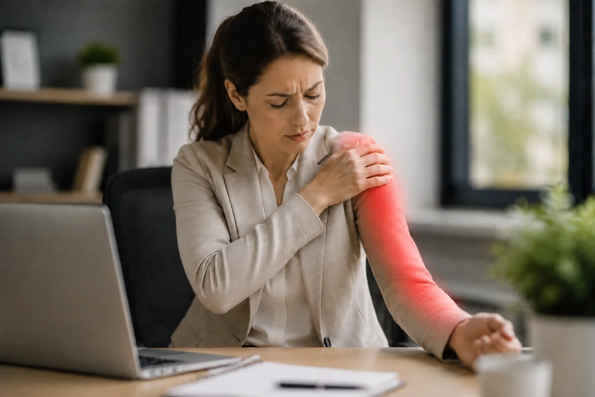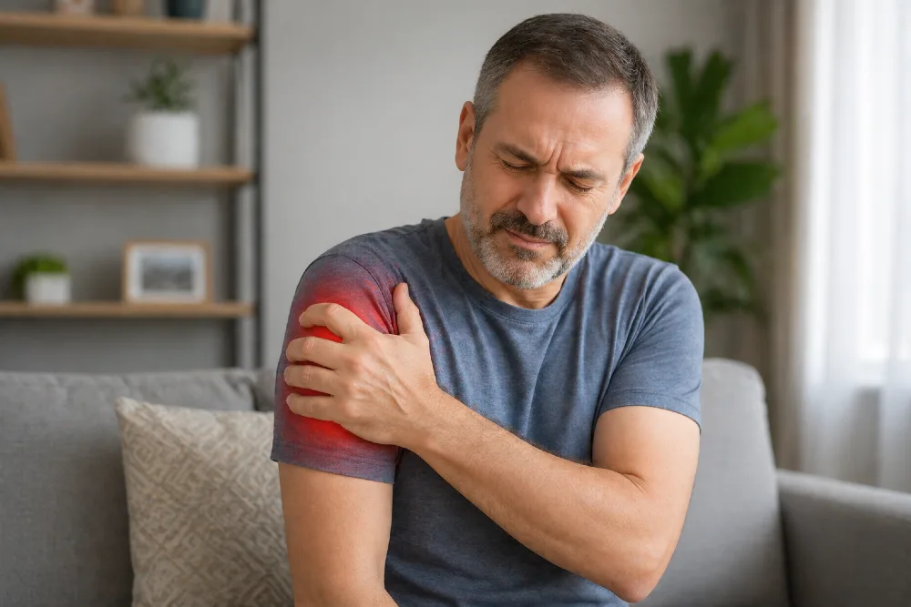
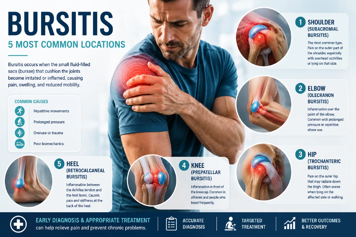
Bursitis is an inflammation or irritation of a bursa — a small fluid-filled sac located between tendons, muscles, and bones. The main function of a bursa is to reduce friction and allow smooth movement around joints. When a bursa becomes irritated, it can lead to localized pain, swelling, tenderness, and reduced movement of the affected area.
Bursitis most commonly affects the shoulder (subacromial bursitis), elbow (olecranon bursitis), hip (trochanteric bursitis), knee (prepatellar or infrapatellar bursitis), and heel. Symptoms depend on the location of the affected bursa but typically include pain with pressure or movement, tenderness, swelling, and sometimes redness or warmth of the skin.
This article explains the most common types of bursitis, why they develop, how to recognize the symptoms, how the condition is diagnosed, and the most effective treatment and prevention strategies.
Key Clinical Points – Bursitis
- Bursitis is inflammation of a bursa, a small fluid-filled sac that reduces friction between bones, tendons, muscles, and skin during movement.
- The most common locations of bursitis include the elbow (olecranon bursitis), knee bursitis, hip bursitis (greater trochanteric pain syndrome), shoulder bursitis, and heel bursitis.
- Repetitive pressure, friction, and overuse are the most common causes, although bursitis can also result from trauma, infection, gout, or inflammatory diseases.
- Typical symptoms include localized pain, tenderness, swelling, and reduced movement. Superficial bursae, such as those around the elbow and knee, may cause visible swelling.
- Diagnosis is usually based on clinical examination, while ultrasound, MRI, or bursal aspiration may be used in selected cases, especially when infection is suspected.
- Most cases improve with conservative treatment including activity modification, ice, medication, and rehabilitation. More persistent cases may require aspiration, injections, or rarely surgery.
What Are Bursae?
A bursa (plural: bursae) is a small, thin, fluid-filled sac that helps reduce friction between moving tissues. It is filled with a small amount of synovial fluid and lined with a synovial membrane, similar to the inner lining of a joint.
Bursae are located between bones and soft tissues such as tendons, muscles, and skin. When healthy, a bursa contains only a very thin layer of fluid with a consistency similar to egg white, allowing smooth gliding between surrounding structures.
The main function of bursae is to decrease friction and facilitate movement around joints. The human body contains more than 150 bursae, with many of the most important ones located around large joints such as the shoulder, hip, knee, and elbow.
When a bursa becomes irritated or inflamed, it can enlarge and become painful, leading to a condition known as bursitis.
What Is Bursitis?
Bursitis refers to inflammation or irritation of a bursa. When a bursa becomes affected by an inflammatory process, its walls may become thickened and the sac can fill with excess fluid.
This increased fluid and thickening of the bursa can cause localized pain, tenderness, and swelling in the affected area. Depending on its location, an enlarged bursa may also be visible as a soft lump beneath the skin, especially in areas such as the elbow (olecranon bursitis) or knee (prepatellar bursitis).
Bursitis pain is usually located directly over the affected bursa and may worsen with pressure, movement, or activities that repeatedly irritate the area. The severity of symptoms depends on the underlying cause and the location of the involved bursa.
What Causes Bursitis?
Bursitis most commonly develops due to prolonged pressure on a bursa or repetitive movements that cause friction between the bursa and surrounding tissues. Over time, this repeated irritation can lead to thickening of the bursal wall, increased fluid production, and pain.
Typical examples include leaning the elbows on hard surfaces, which may contribute to olecranon bursitis, or frequent kneeling without protection, which can lead to prepatellar bursitis. These repeated mechanical stresses gradually irritate the bursa and trigger symptoms.
Bursitis can also occur after direct trauma, such as a blow or fall, or due to infection (septic bursitis). Certain systemic conditions, including rheumatoid arthritis, gout, and lupus, can increase the risk of developing bursitis.
In some cases, the exact cause cannot be identified. Septic bursitis, however, requires prompt medical evaluation because infection can spread and lead to more serious complications.
Who Is Most Likely to Develop Bursitis?
Bursitis can affect people of all ages and occurs in both men and women. However, certain groups have a higher risk depending on their activities, occupation, and other health factors.
Some forms of bursitis are more common in women, particularly trochanteric bursitis and pes anserine bursitis of the knee. These conditions are also more frequently seen in people with increased body weight, which may increase mechanical stress around the hip and knee.
Men are more commonly affected by olecranon bursitis, partly because of higher exposure to occupations involving repetitive elbow pressure and heavy manual work, such as construction, plumbing, gardening, and mechanical work.
People at increased risk of developing bursitis include those who:
- perform repetitive movements involving a specific joint
- have jobs that require prolonged kneeling, leaning, or pressure on joints
- participate in sports with repetitive loading of certain body regions
- have inflammatory conditions such as rheumatoid arthritis or gout
- have previous injuries around a bursa
Although some types of bursitis are associated with specific activities or professions, the underlying mechanism is usually repeated mechanical stress that exceeds the ability of the bursa to adapt.
The Most Common Locations of Bursitis
Although bursitis can develop in many areas of the body, it most commonly affects regions where bursae are exposed to repeated pressure, friction, or mechanical stress. The location of symptoms depends on which bursa is irritated, and each type of bursitis has specific causes, symptoms, and treatment considerations.
The most frequently affected areas include the shoulder, hip, knee, elbow, and heel. Understanding the differences between these conditions is important because pain around a joint does not always originate from the bursa itself and may be caused by other structures such as tendons, ligaments, or nerves.
In the following sections, we will describe the most common types of bursitis, their typical symptoms, and the factors that contribute to their development.
| Location | Common Types of Bursitis | Typical Causes and Features |
|---|---|---|
| Elbow | Olecranon bursitis | The most common bursitis. Often caused by prolonged pressure on the elbow, repetitive irritation, trauma, or infection. Commonly known as “student’s elbow” or “plumber’s elbow”. |
| Knee | Prepatellar bursitis, infrapatellar bursitis, pes anserine bursitis | Frequently related to prolonged kneeling, repetitive stress, or overuse. Prepatellar bursitis is known as “housemaid’s knee”, while infrapatellar bursitis is called “clergyman’s knee”. |
| Hip | Trochanteric bursitis (greater trochanteric bursitis) | Causes pain on the outer side of the hip. Common in runners, people with prolonged pressure on the hip, women, and individuals with excess body weight. |
| Shoulder | Subacromial bursitis | Usually associated with repetitive overhead activities, rotator cuff disorders, trauma, calcific tendinitis, or inflammatory diseases. Causes pain during arm elevation. |
| Heel | Retrocalcaneal bursitis, superficial calcaneal bursitis | Caused by irritation between the Achilles tendon and heel bone or pressure from footwear. Common in runners, dancers, and athletes. |
| Foot | Intermetatarsal bursitis, bunion-associated bursitis | Often related to footwear pressure, foot deformities, and repetitive loading during walking or sports activities. |
| Hip / Pelvic Region | Ischial bursitis | Inflammation of the bursa over the sitting bones. Associated with prolonged sitting and repetitive pressure, sometimes called “weaver’s bottom”. |
| Other Locations | Bursitis around the wrist, hand, ankle, and other joints | Less common and usually related to repetitive pressure, trauma, inflammatory diseases, or infection. |
1. Elbow Bursitis (Olecranon Bursitis)
Elbow bursitis, also known as olecranon bursitis, is a condition characterized by inflammation and swelling of the bursa located at the back of the elbow. The name comes from the olecranon, the bony prominence that forms the pointed tip of the elbow and can be easily felt when the elbow is bent. Olecranon bursitis is one of the most common forms of bursitis.
It most often develops due to prolonged pressure or repetitive irritation of the bursa, especially in people who frequently lean on their elbows on hard surfaces. This is commonly seen in occupations that involve prolonged elbow pressure, such as plumbing, air conditioning installation, mechanical work, and other manual jobs. Because of its association with certain activities, olecranon bursitis is sometimes referred to as “student’s elbow” or “barman’s elbow.” It can also occur after a direct blow to the elbow or in athletes who repeatedly place pressure on the elbows, such as during plank exercises.
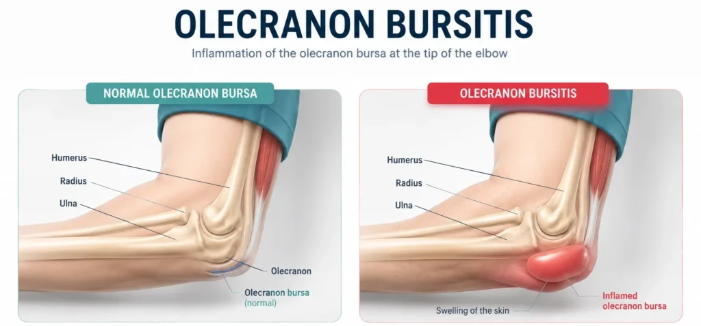
The most common symptom of elbow bursitis is a visible swelling at the back of the elbow, which may appear suddenly after an injury or gradually over time. The size of the swelling can vary significantly. Some people experience only mild discomfort, while others may develop tenderness, redness, and warmth of the skin, especially when the bursa becomes infected (septic bursitis).
Treatment of olecranon bursitis is usually conservative and includes avoiding pressure on the elbow, cold therapy, compression, and pain-relieving medication when needed. In many cases, symptoms improve within several weeks. More persistent or severe cases may require bursal aspiration, corticosteroid injection in selected situations, or rarely surgical removal of the bursa, particularly when recurrent or infected bursitis does not respond to other treatments.
Learn more about the causes, symptoms, diagnosis, and treatment options for this condition in our detailed guide: Elbow Bursitis (Olecranon Bursitis): Symptoms, Causes, and Treatment .
2. Knee Bursitis
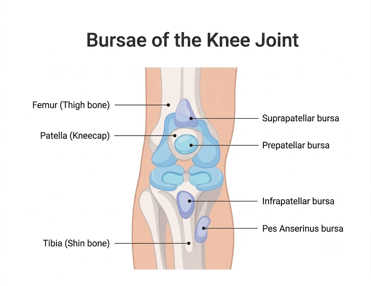
Knee bursitis refers to inflammation of one or more bursae located around the knee joint. The frequency of this condition depends on factors such as age, sex, occupation, physical activity, and individual biomechanics. Several bursae are found around the knee, but the most commonly affected are the prepatellar bursa, pes anserine bursa, and infrapatellar bursa.
The symptoms and causes vary depending on the location of the affected bursa, but typical features include localized pain, swelling, tenderness, and discomfort with movement or direct pressure.
Prepatellar Bursitis
Prepatellar bursitis develops when the bursa located directly in front of the patella (kneecap), between the skin and the patella, becomes irritated and inflamed. It is the most common type of knee bursitis and one of the most frequent forms of bursitis overall, after olecranon bursitis.
It commonly affects people who spend long periods kneeling, such as floor installers, carpenters, gardeners, and construction workers. Because of this association, it is often called “housemaid’s knee.”
The main symptom is swelling at the front of the knee, which may range from a small lump to a larger fluid-filled swelling. Pain is usually worse with kneeling or direct pressure on the area. Redness, warmth, and increased tenderness may occur if the bursa becomes infected (septic bursitis).
Pes Anserine Bursitis
Pes anserine bursitis affects the bursa located on the inner side of the knee, several centimeters below the joint line. It is situated beneath the attachment of the tendons of three muscles that connect the thigh muscles to the upper part of the tibia.
This type of bursitis is more common in middle-aged and older women, particularly those with increased body weight or altered knee alignment, such as valgus knee deformity.
Typical symptoms include pain and tenderness on the inner side of the knee, especially when climbing stairs, walking uphill, or rising from a chair. Swelling is usually mild compared with prepatellar bursitis.
Infrapatellar Bursitis
Infrapatellar bursitis occurs below the patella, where the bursa lies between the patellar tendon and the tibia. It may develop due to repetitive stress, frequent kneeling, or activities that place increased pressure on the front of the knee.
Because of its association with prolonged kneeling, it is sometimes referred to as “clergyman’s knee.”
Symptoms include localized pain and swelling below the kneecap, which may worsen during kneeling, squatting, climbing stairs, or other activities that increase pressure on the area.
Clinical Insight
Each type of knee bursitis requires an individualized treatment approach. Early recognition of symptoms, avoiding activities that repeatedly irritate the bursa, and appropriate rehabilitation are important for successful recovery.
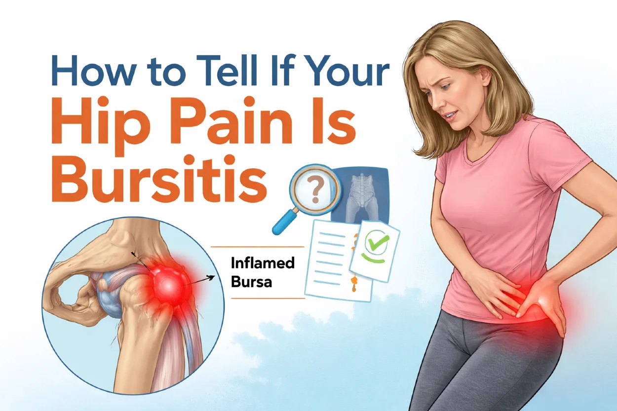
3. Hip Bursitis (Trochanteric Bursitis)
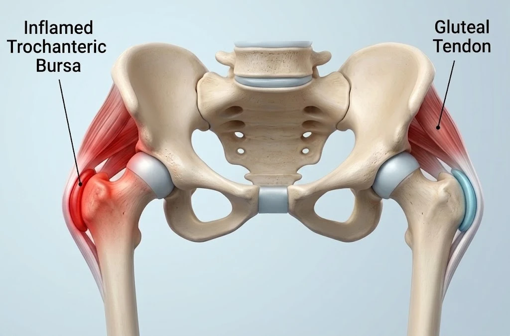
Hip bursitis most commonly refers to irritation of the trochanteric bursa, located on the outer side of the hip, above the bony prominence of the femur called the greater trochanter. This bursa normally helps reduce friction between the greater trochanter and the surrounding soft tissues, including the gluteal tendons, during hip movement.
However, lateral hip pain is often not caused by isolated bursitis alone. In many patients, the condition is part of a broader disorder known as greater trochanteric pain syndrome (GTPS), which includes irritation of the trochanteric bursa together with changes in the gluteus medius and gluteus minimus tendons.
The main symptom is pain on the outer side of the hip, which may spread toward the outer thigh or buttock. Pain is typically aggravated by activities such as walking, climbing stairs, standing up from a chair, or lying on the affected side. Unlike elbow or knee bursitis, swelling of the trochanteric bursa is usually not visible because it is located deep beneath the soft tissues.
Hip bursitis and GTPS may develop due to repetitive mechanical stress, increased pressure on the lateral hip, overuse, or altered movement patterns. It is more common in middle-aged and older women and in people with factors such as increased body weight, reduced hip muscle strength, or differences in leg length, which can increase stress on the lateral hip structures.
Treatment is usually conservative and focuses on reducing irritation while restoring the capacity of the affected tissues. It may include activity modification, avoiding prolonged pressure on the hip, physical therapy, and progressive strengthening exercises. In selected cases with persistent symptoms, corticosteroid injections may provide short-term pain relief, while surgery is rarely required.
4. Shoulder Bursitis (Subacromial Bursitis)
Shoulder bursitis most commonly affects the subacromial bursa, which is located between the rotator cuff tendons and the acromion, the bony projection of the shoulder blade. Its primary function is to reduce friction and allow the rotator cuff tendons to glide smoothly during shoulder movement.
When the subacromial bursa becomes irritated, it can cause pain during overhead activities, reaching behind the back, or lifting the arm away from the body. Many people also experience night pain, especially when lying on the affected shoulder.
Shoulder bursitis may develop as a result of repetitive overhead activities, overuse, direct trauma, age-related degenerative changes, or as part of shoulder impingement syndrome. It may also occur in association with rotator cuff tendinopathy, calcific tendinitis of the shoulder, or inflammatory conditions such as rheumatoid arthritis and gout.
Unlike elbow bursitis, the inflamed bursa lies deep beneath the muscles and does not produce visible swelling. Diagnosis is based on the patient’s history and physical examination and is often confirmed with ultrasound, while MRI may be helpful in selected cases, particularly when a rotator cuff tear or other shoulder pathology is suspected.
Treatment is usually non-surgical and includes activity modification, ice application, pain-relieving medication, and physical therapy aimed at restoring normal shoulder mechanics. In persistent cases, an ultrasound-guided corticosteroid injection into the subacromial bursa may provide symptom relief, while surgery is rarely necessary.
5. Heel Bursitis
Heel bursitis most commonly refers to retrocalcaneal bursitis, an inflammation of the bursa located between the Achilles tendon and the heel bone (calcaneus). This bursa normally reduces friction as the Achilles tendon glides over the calcaneus during walking, running, and standing on the toes.
When the bursa becomes inflamed, it causes pain at the back of the heel, often accompanied by tenderness and swelling just above the heel bone. Symptoms typically worsen during activities that place increased strain on the Achilles tendon, such as walking uphill, running, or climbing stairs. Many patients also notice pain during the first few steps in the morning or after prolonged periods of rest.
Heel bursitis commonly affects runners, dancers, and other physically active individuals. It may develop due to repetitive overuse, direct pressure from tight or poorly fitting footwear, or underlying conditions such as Haglund’s deformity, which increases friction between the Achilles tendon and the bursa.
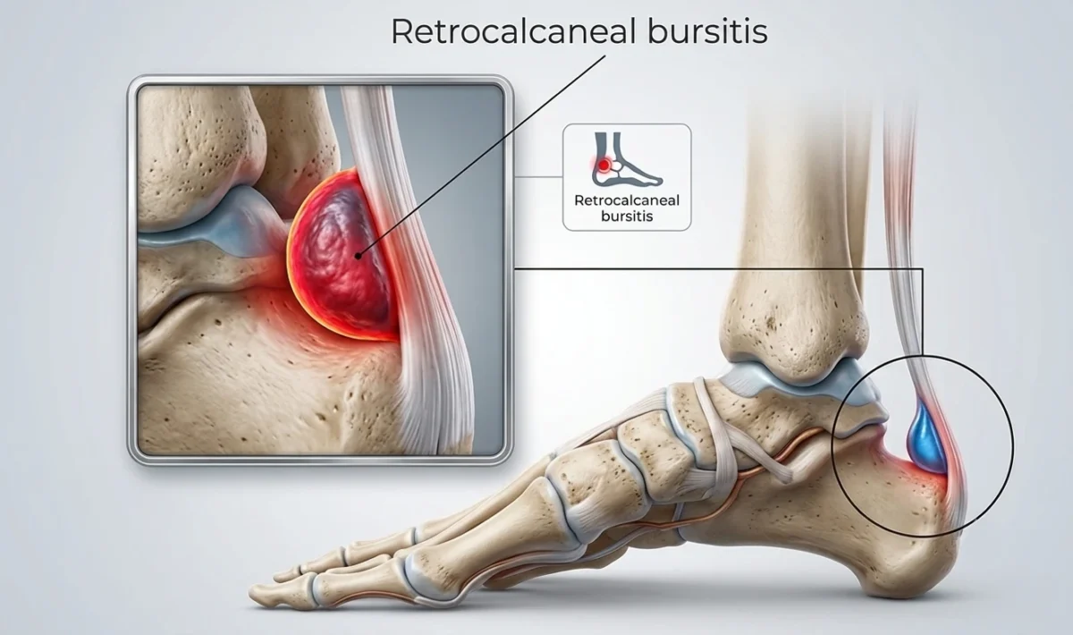
Diagnosis is usually based on the patient’s history, physical examination, and ultrasound, which can visualize the inflamed bursa and assess the Achilles tendon. MRI may be recommended if additional tendon injury or other heel pathology is suspected.
Treatment is usually conservative and includes activity modification, changing footwear, ice application, pain-relieving medication, and physical therapy. In selected cases, corticosteroid injections may be considered with great caution because of the nearby Achilles tendon, while surgery is reserved for patients with persistent symptoms that do not respond to non-surgical treatment.
Symptoms of Bursitis
The symptoms of bursitis vary depending on which bursa is affected, but most patients experience a combination of localized pain, tenderness, swelling, and reduced range of motion. Symptoms may develop gradually due to repetitive stress or appear suddenly after prolonged pressure or minor trauma.
Pain is usually worse during movements that compress or irritate the inflamed bursa and often improves with rest. The affected area may also feel tender when touched.
When superficial bursae, such as those around the elbow or knee, are involved, swelling is often clearly visible as a soft lump beneath the skin. In contrast, inflammation of deeper bursae, such as those around the hip or shoulder, usually does not produce visible swelling and may instead present as poorly localized joint pain that can mimic tendon or muscle disorders.
Common symptoms of bursitis include:
- Localized pain around the affected joint
- Swelling over the inflamed bursa (particularly in superficial bursae)
- Tenderness when pressing on the affected area
- Pain with movement or prolonged pressure
- Reduced range of motion due to pain
Septic Bursitis
Septic bursitis occurs when a bursa becomes infected, most commonly by bacteria entering through a small cut, abrasion, or puncture wound near the affected joint. It most frequently affects superficial bursae, particularly the olecranon bursa at the elbow and the prepatellar bursa at the knee.
Unlike non-infectious bursitis, septic bursitis typically causes rapidly increasing swelling, redness, warmth, severe tenderness, and sometimes fever or chills. Because the infection can spread to surrounding tissues or the bloodstream, prompt medical evaluation is essential.
Treatment usually involves antibiotics, and in many cases the fluid inside the bursa must be aspirated to confirm the diagnosis and relieve pressure. Surgery is reserved for persistent or severe infections that do not respond to conservative treatment.
Diagnosis of Bursitis
The diagnosis of bursitis is primarily based on a clinical examination and a detailed assessment of the patient’s symptoms. During the examination, the doctor evaluates the location of pain, tenderness, swelling, range of motion, and possible signs of infection.
A detailed medical history is also important, including information about occupation, sports activities, repetitive movements, previous injuries, and underlying inflammatory conditions such as rheumatoid arthritis or gout. These factors can help identify the cause of bursitis and distinguish it from other conditions that may cause similar symptoms.
Additional tests are not always required but may be recommended when the diagnosis is uncertain, symptoms persist, or infection is suspected. These may include:
- Ultrasound examination – commonly used because it can quickly visualize fluid accumulation, thickening of the bursa, and inflammation in real time.
- Magnetic resonance imaging (MRI) – may be useful for deeper bursae, complex cases, or when other soft tissue problems are suspected.
- Bursal aspiration – removal of fluid from the inflamed bursa for laboratory analysis, especially when septic bursitis, gout, or other inflammatory conditions are suspected.
- Blood tests – inflammatory markers such as CRP, ESR, and white blood cell count may support the evaluation when infection or systemic inflammation is suspected.
Accurate diagnosis is important because pain around a joint is not always caused by bursitis and may originate from nearby tendons, ligaments, muscles, nerves, or the joint itself.
Bursitis Treatment
Most cases of bursitis can be successfully treated with conservative (non-surgical) methods. The main goals of treatment are to reduce irritation of the affected bursa, relieve pain, and restore normal function.
The first step is usually activity modification and avoiding movements or positions that repeatedly aggravate symptoms. This does not always require complete rest, but reducing pressure and repetitive stress on the affected area allows the bursa to recover. Cold therapy may help decrease pain and swelling, while non-steroidal anti-inflammatory drugs (NSAIDs), such as ibuprofen or diclofenac, may provide short-term symptom relief.
Physical therapy can be beneficial, especially when bursitis is related to repetitive loading, muscle weakness, altered movement patterns, or biomechanical problems. Rehabilitation focuses on improving mobility, restoring muscle function, and reducing stress on the affected tissues.
If symptoms do not improve with conservative treatment, additional procedures may be considered. Bursal aspiration (removal of excess fluid with a needle) may be performed when there is significant swelling, discomfort, or suspicion of infection. Corticosteroid injections into the bursa can provide short-term pain relief in selected patients, but they should be used cautiously because repeated injections may negatively affect surrounding tissues.
In cases of septic bursitis, treatment requires antibiotic therapy, and aspiration is often necessary to analyze the fluid and guide treatment. Surgery, such as bursectomy (removal of the inflamed bursa), is rarely needed and is generally reserved for persistent, recurrent cases or infections that do not respond to other treatments.
Prevention of Bursitis
Although bursitis cannot always be completely prevented, especially in people with inflammatory conditions or certain anatomical factors, many cases can be avoided by reducing repeated stress on joints and bursae.
The key to prevention is minimizing prolonged pressure, repetitive friction, and excessive loading of vulnerable areas. This can be achieved through simple adjustments in daily activities, work habits, and exercise routines.
Important prevention strategies include:
- Using protective padding during activities that place pressure on joints, such as knee pads for prolonged kneeling or soft supports when leaning on the elbows.
- Improving workplace ergonomics by adjusting positions, tools, and working techniques to reduce repetitive stress.
- Avoiding prolonged pressure on specific body areas, especially the elbows, knees, and hips.
- Maintaining muscle strength and flexibility, particularly around major joints such as the shoulder, hip, and knee, to improve movement mechanics and reduce unnecessary stress on bursae.
- Warming up properly before exercise and gradually increasing training intensity to allow tissues to adapt.
- Using appropriate footwear to reduce pressure and friction around the heel and Achilles tendon.
At the first signs of irritation, such as localized pain, swelling, or tenderness, it is important to modify activities and address symptoms early. Timely management can help prevent progression to chronic bursitis and reduce the risk of recurrent symptoms.
Frequently Asked Questions About Bursitis
What is bursitis?
Bursitis is inflammation of a bursa, a small fluid-filled sac that reduces friction between bones, tendons, muscles, and skin. When a bursa becomes irritated, it may become swollen and painful, causing localized discomfort and reduced movement.
What causes bursitis?
Bursitis most commonly develops due to repetitive pressure, friction, or overuse of a specific area. Activities such as prolonged kneeling, leaning on the elbows, repetitive shoulder movements, or excessive loading during sports can irritate a bursa. Trauma, infection, and inflammatory conditions such as gout or rheumatoid arthritis can also cause bursitis.
How long does bursitis take to heal?
The recovery time depends on the affected bursa, severity of inflammation, and whether the underlying cause is addressed. Mild cases often improve within a few weeks with activity modification and appropriate treatment. Chronic or recurrent bursitis may require a longer rehabilitation period.
Can bursitis go away on its own?
Mild cases of bursitis may improve with rest, avoiding activities that aggravate symptoms, and reducing pressure on the affected area. However, persistent pain, swelling, or recurrent symptoms may require medical evaluation to identify the cause and determine the most appropriate treatment.
What is the best treatment for bursitis?
Treatment depends on the type and severity of bursitis but usually includes activity modification, ice application, pain-relieving medication, and physical therapy. In some cases, procedures such as bursal aspiration or corticosteroid injections may be considered. Septic bursitis requires antibiotic treatment.
Can bursitis come back after treatment?
Yes, bursitis can recur, especially if the activity or mechanical stress that caused it continues. Preventing recurrence involves modifying provoking activities, improving ergonomics, strengthening surrounding muscles, and avoiding prolonged pressure on the affected area.
What is the difference between bursitis and tendonitis?
Bursitis affects a bursa, which is a fluid-filled sac that reduces friction between tissues. Tendonitis (or more accurately, tendinopathy in many chronic cases) affects a tendon, the structure that connects muscle to bone. Both conditions can cause pain around a joint and may occur together.
When should I see a doctor for bursitis?
You should seek medical advice if you have severe pain, rapidly increasing swelling, redness, warmth, fever, or difficulty moving the affected joint. These symptoms may indicate septic bursitis or another condition requiring specific treatment.
Williams CH, Jamal Z, Sternard BT. Bursitis. StatPearls [Internet]. Treasure Island (FL): StatPearls Publishing; 2025 Jan. Evidence-based overview: Bursitis causes, diagnosis, and treatment
InformedHealth.org (IQWiG). Overview: Bursitis. Institute for Quality and Efficiency in Health Care. Patient-oriented review: Symptoms, causes, and treatment of bursitis
Synovial Bursa. Wikipedia. Anatomical overview: Structure and function of synovial bursae
Rishor-Olney CR, Taqi M, Pozun A. Prepatellar Bursitis. StatPearls [Internet]. Treasure Island (FL): StatPearls Publishing; 2025 Jan. Clinical review: Diagnosis and management of prepatellar bursitis
Chatra PS. Bursae around the knee joints. Indian Journal of Radiology and Imaging. 2012;22(1):27-30. Imaging reference: Anatomy and ultrasound appearance of knee bursae
Seidman AJ, Taqi M, Varacallo MA. Trochanteric Bursitis. StatPearls [Internet]. Treasure Island (FL): StatPearls Publishing; 2025 Jan. Clinical review: Greater trochanteric bursitis diagnosis and treatment