
Gout, also known as gouty arthritis, is an inflammatory form of arthritis caused by the buildup of monosodium urate crystals in and around the joints. The condition has been recognized since ancient times and was historically known as the “disease of kings” because of its association with rich food and alcohol. Although certain dietary and lifestyle factors can increase the risk of gout, genetics and kidney function also play an important role.
Gout is the most common form of inflammatory arthritis. It can affect people of different ages, but it is particularly common in middle-aged and older men. A typical gout attack develops suddenly, causing intense joint pain, swelling, redness, and warmth. The big toe is the most characteristic location, a presentation known as podagra, although gout can also affect the ankle, knee, foot, and other joints.
Without appropriate treatment, recurrent gout attacks can eventually lead to chronic gout, the formation of tophi, and permanent joint damage. The good news is that gout can usually be effectively managed by treating acute attacks and, when indicated, lowering uric acid levels to prevent future attacks and complications.
In this article, we explain what causes gout, how to recognize its symptoms, how gout is diagnosed and treated, and what you can do to prevent future attacks.
What Is Gout and What Causes It?
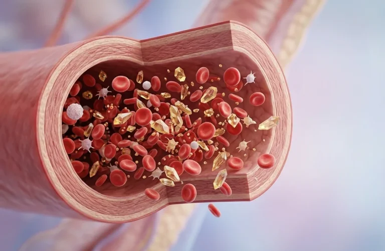
Gout, also known as gouty arthritis, is an inflammatory joint disease caused by the deposition of monosodium urate crystals in and around the joints. Uric acid is produced when the body breaks down substances called purines and is normally present in the blood at low concentrations. When blood uric acid levels become persistently elevated—a condition known as hyperuricemia—the risk of developing gout increases.
Urate normally remains dissolved in the blood. When its concentration exceeds the level at which it can remain soluble, monosodium urate crystals can begin to form and accumulate in joints and surrounding tissues. These crystals trigger a strong immune response, activating inflammatory cells and mediators. This produces the characteristic pain, swelling, redness, and warmth of an acute gout attack.
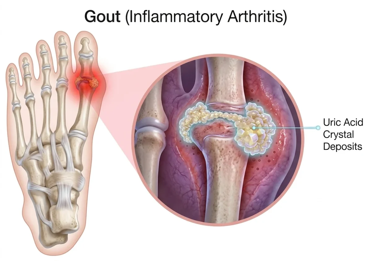
Over time, continued urate crystal deposition can lead to the formation of tophi—collections of urate crystals that develop in and around joints and other tissues. Tophi can become visible or palpable under the skin and, in advanced gout, may contribute to joint destruction and functional impairment.
Hyperuricemia can develop for several reasons. In most people with gout, the main problem is reduced elimination of urate by the kidneys, although increased urate production can also contribute. Diets rich in purines, alcohol consumption, obesity, kidney disease, certain medications, and genetic factors can all influence uric acid levels and the risk of gout.
Importantly, not everyone with hyperuricemia develops gout. Elevated uric acid increases the likelihood of crystal formation and future gout attacks, but many people with hyperuricemia remain asymptomatic.
Gout can occur as intermittent acute attacks, often separated by symptom-free periods, or progress to chronic gout with persistent crystal deposition, tophi, recurrent inflammation, and joint damage. The next sections examine the symptoms and clinical features of gout, followed by the major risk factors that increase the likelihood of developing the disease and experiencing recurrent attacks.
Gout Through History
Gout has been recognized for centuries, with descriptions of its characteristic attacks appearing in ancient Egyptian medical records and the writings of Hippocrates. For much of history, it became associated with wealth and excess and was famously called the “disease of kings” or the “disease of the rich.”
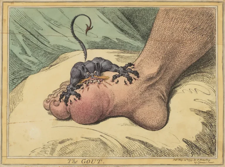
This reputation was largely related to the diets and lifestyles of wealthy people, which often included abundant meat and alcohol. Gout was also depicted in art as a symbol of its excruciating pain. One of the best-known examples is James Gillray’s 1799 satirical print The Gout, which shows a large claw-like creature gripping the sufferer’s foot.
Several well-known historical figures, including Henry VIII and Benjamin Franklin, are believed to have suffered from gout, further reinforcing its association with wealth and indulgence.
Today, however, we understand that gout is far more complex than a disease caused simply by rich food or alcohol. Diet and lifestyle can influence uric acid levels and trigger attacks, but genetic factors, kidney function, and other medical conditions play an important role in determining who develops gout.

Risk Factors for Gout
Gout usually develops as a result of a combination of genetic predisposition, medical conditions, medications, and lifestyle factors. Some risk factors cannot be changed, while others can be modified to reduce the likelihood of developing gout or experiencing recurrent attacks.
Non-Modifiable Risk Factors
Important factors that cannot be changed include:
- Age: Gout becomes more common with increasing age.
- Sex: It is more common in men, while the risk in women increases after menopause.
- Family history: Having close relatives with gout increases the risk because genetic factors influence uric acid metabolism and kidney excretion.
- Genetic predisposition: Certain genetic variations can affect how efficiently the body eliminates uric acid.
Modifiable Risk Factors
Several health conditions and lifestyle factors can increase uric acid levels or make gout attacks more likely:
- Obesity and metabolic disorders, including hypertension and diabetes.
- Chronic kidney disease, which can reduce uric acid elimination.
- Certain medications, particularly diuretics, which may increase uric acid levels.
- Alcohol consumption, especially beer and spirits.
- Purine-rich foods, including organ meats, red meat, and some seafood.
- Sugar-sweetened beverages and high-fructose foods, which can contribute to increased uric acid levels.
Clinical Insight
It is important to remember that diet alone does not explain why someone develops gout. Genetics, kidney function, age, sex, medications, and other health conditions can be equally or more important. Identifying modifiable risk factors can nevertheless help reduce uric acid levels and lower the risk of recurrent gout attacks and long-term complications.
What Triggers a Gout Attack?
A gout attack occurs when monosodium urate crystals trigger an intense inflammatory response inside or around a joint. Although elevated serum urate is the underlying risk factor, certain situations can suddenly disturb urate balance or trigger inflammation and precipitate an attack.
Common gout triggers include:
- Alcohol, particularly beer and spirits
- Large meals and excessive intake of purine-rich foods
- Dehydration, which can contribute to changes in urate concentration
- Fasting or rapid weight loss
- Joint injury or physical stress
- Surgery or acute illness
- Certain medications, particularly diuretics
- Starting or changing urate-lowering therapy, which can temporarily increase the risk of a flare as urate crystals begin to dissolve
It is important to distinguish a trigger from the underlying cause of gout. For example, eating a large meal or drinking alcohol may precipitate an attack, but the development of gout is usually related to a combination of genetic factors, impaired renal urate excretion, and persistent hyperuricemia.
Not every gout attack has an identifiable trigger. Attacks can occur even when a person has not made any obvious dietary or lifestyle changes.
Related Guides

Clinical Manifestations of Gout
Gout can present in several stages, ranging from sudden acute gout attacks to chronic disease with persistent urate crystal deposition and joint damage. Importantly, most people with hyperuricemia never develop symptomatic gout. However, the risk of gout increases as serum urate levels rise.
The clinical course typically includes acute attacks, symptom-free intercritical periods, and, in some patients, chronic gout with recurrent inflammation and tophi.
Acute Gout
An acute gout attack usually develops suddenly, often overnight, with severe pain and inflammation in a single joint. The classic presentation is podagra, an attack affecting the first metatarsophalangeal (MTP) joint at the base of the big toe. The pain can be extremely intense, and even light pressure from a bedsheet may be intolerable.
During an acute attack, the affected joint typically becomes swollen, red, warm, and extremely tender. The inflammation results from the interaction between monosodium urate crystals and the immune system, which triggers a strong inflammatory response.
Several factors can precipitate a gout attack, including:
- Joint injury or physical stress
- Heavy alcohol consumption
- Large meals or dietary changes
- Fasting or rapid weight loss
- Certain medications, particularly diuretics
- Starting or changing urate-lowering therapy
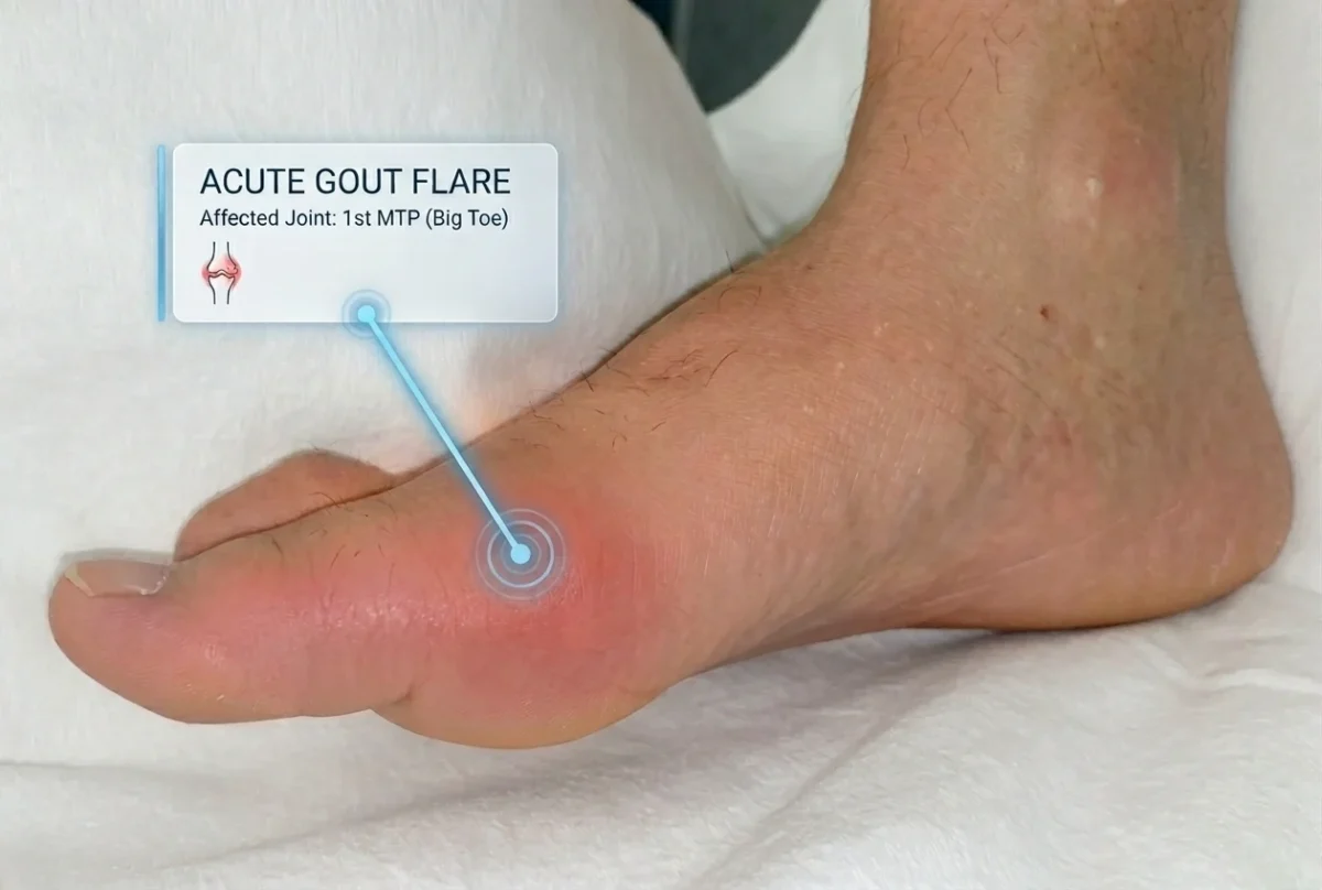
Although the big toe is the most characteristic location, gout can also affect the ankles, knees, wrists, fingers, and other joints. Urate crystal inflammation can also involve surrounding structures such as tendons, bursae, and other soft tissues.
An untreated acute gout attack often lasts several days to a few weeks and eventually subsides, even without treatment. However, recurrent attacks are common when the underlying hyperuricemia remains untreated.
The Intercritical Period
After an acute attack resolves, most people enter an intercritical period, during which the affected joint may return to normal and the patient may have no symptoms.
This symptom-free period does not necessarily mean that the underlying disease has disappeared. If serum urate remains elevated, urate crystals can continue to accumulate in the joints and tissues, increasing the risk of future gout attacks and long-term complications.
Chronic Gout
Without adequate treatment, gout may gradually progress to chronic gout, characterized by recurrent attacks, persistent urate crystal deposition, and progressive joint damage. Over time, some patients develop tophi—collections of monosodium urate crystals that can form in and around joints and other tissues.
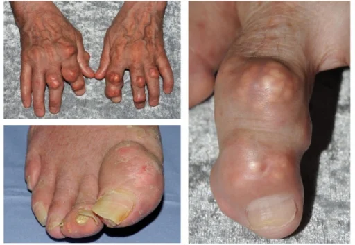
Tophi commonly develop around the fingers, toes, elbows, and ears and may appear as firm or whitish lumps beneath the skin. In advanced cases, they can ulcerate and release a chalky material. Although tophi are characteristic of longstanding gout, they can occasionally appear earlier in the course of the disease.
Chronic tophaceous gout can cause joint destruction, deformity, reduced mobility, and loss of function. Persistent inflammation and crystal deposition can also damage bone and surrounding soft tissues.
Gout is also associated with several conditions affecting other organs. Longstanding hyperuricemia can contribute to uric acid kidney stones, while people with gout commonly have a higher burden of chronic kidney disease and cardiovascular disease. These associations are important when assessing gout as a systemic metabolic condition rather than simply a problem affecting the joints.
How Is Gout Diagnosed?
The diagnosis of gout is based on the clinical presentation, laboratory testing, and, when necessary, analysis of joint fluid or imaging studies. Recognizing gout accurately is important because several other conditions, particularly septic arthritis, can cause a similar hot, swollen, and painful joint.
Clinical Presentation
Gout typically presents with a sudden attack of severe joint pain, often affecting a single joint. The first metatarsophalangeal (MTP) joint of the big toe is the classic location, known as podagra, but the knees, ankles, wrists, and fingers can also be affected.
During an acute attack, the joint is typically swollen, red, warm, and extremely tender. Even minimal movement or light pressure from clothing or bedding may cause significant pain. A typical clinical presentation can strongly suggest gout, but it does not always establish the diagnosis with certainty.
Blood Tests
A serum uric acid test is commonly performed when gout is suspected. However, an elevated uric acid level does not by itself diagnose gout, because many people with hyperuricemia never develop the disease.
Conversely, serum urate can be normal during an acute gout attack. Therefore, a normal result during a flare does not reliably exclude gout, particularly when the clinical presentation is strongly suggestive.
Blood tests may also show evidence of inflammation, such as elevated C-reactive protein (CRP) or erythrocyte sedimentation rate (ESR), and sometimes an increased white blood cell count. These findings indicate inflammation but are not specific to gout.
Joint Aspiration and Crystal Analysis
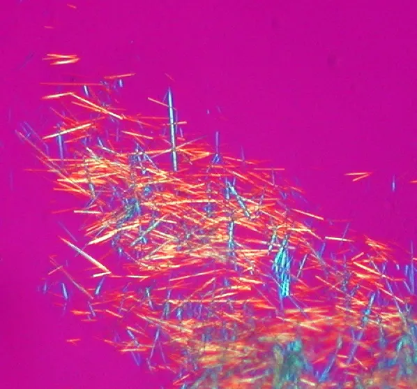
When the diagnosis is uncertain, joint aspiration (arthrocentesis) can provide definitive confirmation. A sample of synovial fluid is removed from the affected joint and examined under polarized light microscopy.
The identification of needle-shaped monosodium urate crystals with strong negative birefringence confirms gout.
Joint aspiration is particularly important when septic arthritis is a possibility, because infection can closely resemble an acute gout attack. Synovial fluid can be analyzed for both crystals and microorganisms to help distinguish these conditions.
Imaging Tests
Imaging can be useful when the diagnosis is uncertain, when joint aspiration is not feasible, or when chronic gout and tophi are suspected.
Ultrasound can detect urate deposits and may show characteristic findings such as the double contour sign over the surface of articular cartilage. It can also identify tophi and joint inflammation and can be used to guide joint aspiration or other procedures.
Dual-energy CT (DECT) can specifically identify and quantify urate deposits and is particularly useful when the diagnosis remains uncertain or when deposits are located in deeper tissues.
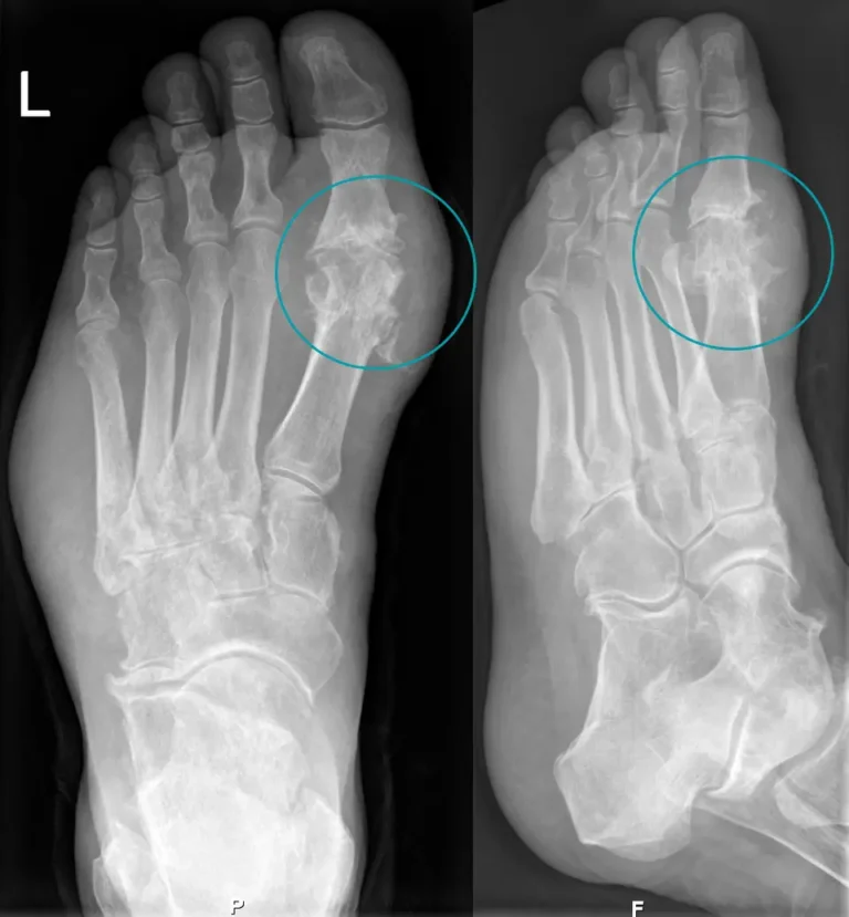
X-rays are less useful during an early acute attack but can show characteristic changes in longstanding gout, including erosions and joint damage. MRI can provide detailed information about soft tissues, bone, and tophi but is not usually the first imaging test for diagnosing gout.
Gout and Uric Acid Levels
Uric acid plays a central role in the development of gout. When serum urate remains elevated, monosodium urate crystals can form and accumulate in joints and surrounding tissues. A level above approximately 6.8 mg/dL (404 µmol/L) exceeds the saturation point at which these crystals can form.
However, high uric acid does not necessarily mean that a person has gout. Many people with hyperuricemia never develop gout, while serum urate can occasionally be normal during an acute gout attack.
For people with established gout, the usual treatment target is less than 6 mg/dL (360 µmol/L). A lower target may be appropriate in patients with severe or tophaceous gout. Maintaining urate below the target level over time helps dissolve existing crystals and prevent future gout attacks and joint damage.
Gout Treatment
The treatment of gout has two main goals: controlling the intense inflammation and pain of an acute gout attack, and lowering uric acid levels over the long term to prevent future attacks and complications.
The main goals of gout treatment are to:
- Relieve pain and inflammation during an acute attack
- Prevent recurrent gout attacks
- Lower serum urate levels when urate-lowering therapy is indicated
- Prevent tophi and chronic gout
- Protect the joints and prevent long-term damage
Treatment of an Acute Gout Attack
Treatment of an acute gout attack should be started as soon as possible, because early treatment can reduce the severity and duration of symptoms. The main medications used are NSAIDs, colchicine, and corticosteroids. The choice depends on the patient’s other medical conditions, medications, kidney function, and previous response to treatment.
NSAIDs such as naproxen, indomethacin, or ibuprofen are commonly used to control the inflammation and severe pain associated with an acute gout attack. They are effective when started early, but they may not be suitable for people with certain kidney, gastrointestinal, or cardiovascular conditions.
Colchicine is another established treatment for acute gout. It works by reducing the inflammatory response to monosodium urate crystals and is most effective when started early in the attack. Because colchicine has a relatively narrow therapeutic range, the dose must be appropriate for the individual patient, particularly in people with kidney or liver disease or those taking interacting medications. Gastrointestinal symptoms, especially diarrhea, are among its most common adverse effects.
Corticosteroids can be used when NSAIDs or colchicine are unsuitable or ineffective. They may be given orally, by injection, or directly into the affected joint when only one or a few joints are involved. Intra-articular corticosteroid injection can provide rapid relief while limiting systemic exposure.
In addition to medication, simple measures can help reduce discomfort during an acute attack. Resting and elevating the affected joint and applying a cold pack for short periods may help reduce pain and swelling. Avoiding pressure on the affected joint can also make walking or other daily activities more tolerable.
Treatment of the acute attack controls the immediate inflammation, but it does not eliminate the underlying cause of gout. When indicated, long-term urate-lowering therapy is needed to reduce the body’s urate burden, prevent recurrent attacks, dissolve existing urate crystals, and reduce the risk of tophi and joint damage.
Long-Term Treatment of Gout and Prevention of Future Attacks
The main goal of long-term gout treatment is to reduce the body’s urate burden and prevent further attacks, tophi, and permanent joint damage. This is achieved through a combination of lifestyle measures and urate-lowering medication when indicated.
The long-term treatment goals are to:
- Lower serum urate to the recommended target
- Prevent recurrent gout attacks
- Dissolve existing urate crystals and reduce or eliminate tophi
- Prevent further joint damage
- Reduce the long-term complications associated with gout
Diet and Lifestyle Changes
Maintaining a healthy body weight is beneficial for people with gout. If you are overweight or obese, gradual weight loss can help reduce serum urate levels and may decrease the frequency of gout attacks. Regular physical activity can support weight management and overall cardiovascular health.
Diet can also influence uric acid levels and the risk of gout attacks. Rather than following an extremely restrictive diet, most people benefit from an overall healthy, balanced eating pattern with moderation of foods and beverages associated with increased gout risk.
Helpful measures include:
- Limiting organ meats and large amounts of red meat
- Moderating certain seafood, particularly shellfish and other high-purine foods
- Limiting beer and spirits, which can increase the risk of gout attacks
- Reducing sugar-sweetened beverages and high-fructose drinks
- Maintaining adequate hydration
- Choosing a balanced diet rich in vegetables, whole grains, and other minimally processed foods
Dietary changes can help lower uric acid and reduce triggers, but diet alone usually cannot lower serum urate enough to treat established gout when urate-lowering therapy is indicated.
Dietary patterns such as the DASH diet have also been associated with lower serum urate levels and may be a useful approach for people with gout, particularly when combined with overall cardiovascular risk reduction.
Urate-Lowering Therapy
For people who require long-term treatment, the cornerstone of gout management is urate-lowering therapy (ULT). The aim is to maintain serum urate below the level at which urate crystals can persist and gradually dissolve existing deposits.
The 2020 American College of Rheumatology guideline recommends a treat-to-target approach, with regular monitoring and adjustment of medication to achieve a serum urate level of less than 6 mg/dL (360 µmol/L). A lower target may be considered in some patients with severe or tophaceous gout.
The main medications used to lower urate include:
- Allopurinol: A xanthine oxidase inhibitor that reduces uric acid production. It is generally the preferred first-line urate-lowering medication, including for many patients with chronic kidney disease. Treatment is usually started at a low dose and gradually increased according to serum urate levels and the patient’s clinical situation.
- Febuxostat: Another xanthine oxidase inhibitor that reduces uric acid production. It may be used when allopurinol is not tolerated, is contraindicated, or does not adequately control serum urate.
- Uricosuric medications: These increase the renal excretion of uric acid and may be considered in selected patients. Their use is more limited, particularly in people with kidney stones or significant chronic kidney disease.
Importantly, urate-lowering therapy is not routinely recommended for people who have asymptomatic hyperuricemia alone, meaning elevated serum urate without clinical gout.
For patients with established gout, long-term urate-lowering therapy is generally considered when there are recurrent gout attacks, tophi, gout-related joint damage, or certain other high-risk clinical features. The decision should be individualized according to the patient’s medical history and risk factors.
Preventing Gout Attacks When Starting Urate-Lowering Therapy
Starting urate-lowering therapy can temporarily increase the risk of an acute gout attack as urate crystals begin to dissolve and the body’s urate balance changes.
For this reason, guidelines recommend anti-inflammatory prophylaxis when initiating ULT. Depending on the patient’s circumstances, this may include low-dose colchicine, an NSAID, or another appropriate anti-inflammatory medication.
Prophylaxis is generally continued for at least 3–6 months, with longer treatment considered in patients who continue to have gout flares or have persistent tophi.
Clinical Insight
Long-term control of gout therefore involves more than simply treating individual attacks. Consistently lowering serum urate to the target range allows existing urate crystals to dissolve over time and reduces the risk of future attacks, tophi, and joint damage.
Patient Advice
If you have gout, one of the most important things you can do is keep your uric acid level under control, even when you are feeling completely well. Gout often has long symptom-free periods, but urate crystals can continue to accumulate if the underlying hyperuricemia is not treated.
Try to maintain a healthy weight, stay well hydrated, and limit alcohol—particularly beer and spirits—as well as sugary drinks and large amounts of red meat and seafood. You do not need to follow an extremely restrictive diet; a balanced, healthy diet is usually a more sustainable approach.
If your doctor has prescribed urate-lowering medication, take it regularly as directed and do not stop it simply because your joint pain has disappeared. Regular monitoring of your serum urate level helps ensure that treatment is actually reaching the recommended target.
During an acute attack, give the affected joint some rest and avoid unnecessary pressure or strenuous activity. Once the attack settles, gradually return to normal movement and physical activity.
Most importantly, remember that gout is a treatable chronic disease. With consistent treatment and appropriate lifestyle changes, the frequency of attacks can be greatly reduced and long-term joint damage can often be prevented.
Key Clinical Points – Gout
- Gout is an inflammatory arthritis caused by the deposition of monosodium urate crystals in and around joints.
- The classic presentation is podagra — a sudden, extremely painful attack affecting the joint at the base of the big toe.
- Gout can also affect the ankles, knees, wrists, and fingers, causing severe pain, swelling, redness, warmth, and tenderness.
- Hyperuricemia is the main risk factor, but elevated uric acid alone does not mean that a person has gout.
- Diagnosis can be confirmed by identifying monosodium urate crystals in joint fluid; ultrasound and dual-energy CT can also help detect urate deposits.
- Acute attacks are treated with NSAIDs, colchicine, or corticosteroids, depending on the patient’s individual circumstances.
- Long-term treatment aims to maintain serum urate at less than 6 mg/dL (360 µmol/L) to dissolve urate crystals and prevent recurrent attacks, tophi, and joint damage.
- With appropriate urate-lowering treatment and lifestyle changes, gout can usually be effectively controlled and long-term complications prevented.
Frequently Asked Questions About Gout
What is gout?
Gout is an inflammatory form of arthritis caused by the accumulation of monosodium urate crystals in and around joints.
What causes gout?
Gout develops when serum urate remains elevated over time, allowing urate crystals to form. Genetics, kidney function, age, sex, medications, and metabolic conditions can all contribute.
What is podagra?
Podagra is an acute gout attack affecting the first metatarsophalangeal joint at the base of the big toe. It is the classic presentation of gout.
What does a gout attack feel like?
A gout attack usually causes sudden, severe joint pain, accompanied by swelling, redness, warmth, and extreme tenderness. Attacks often develop rapidly, sometimes overnight.
Can gout go away on its own?
An acute gout attack can eventually resolve without treatment, but the underlying urate crystal deposits remain if serum urate is not adequately controlled. Without appropriate treatment, attacks may become more frequent and chronic gout can develop.
What uric acid level causes gout?
There is no single uric acid level that guarantees a person will develop gout. However, persistent serum urate above approximately 6.8 mg/dL (404 µmol/L) increases the likelihood of urate crystal formation.
Can you have gout with normal uric acid?
Yes. Serum urate can be normal during an acute gout attack, so a single normal blood test does not exclude gout.
What is the target uric acid level for gout?
For most people with gout receiving urate-lowering therapy, the target is below 6 mg/dL (360 µmol/L). A lower target may be considered in patients with severe or tophaceous gout.
What foods should you avoid with gout?
It is generally recommended to limit organ meats, large amounts of red meat, certain seafood, alcohol—especially beer and spirits—and sugar-sweetened beverages. A balanced diet is preferable to an extremely restrictive diet.
Is gout a serious disease?
Gout can become serious when it is untreated or poorly controlled. Recurrent attacks can lead to tophi, chronic inflammation, joint damage, and reduced function, while gout is also associated with kidney and cardiovascular disease.
Afzal M, Rednam M, Gujarathi R, et al. Gout. StatPearls [Internet]. Updated 2025 Jun 23. Evidence-based clinical review: Gout symptoms, diagnosis, and management
FitzGerald JD, Dalbeth N, Mikuls T, et al. 2020 American College of Rheumatology Guideline for the Management of Gout. Arthritis Care Res (Hoboken). 2020;72(6):744–760. ACR clinical guideline: Gout treatment and urate-lowering therapy
Shekelle PG, FitzGerald J, Newberry SJ, et al. Management of Gout. Agency for Healthcare Research and Quality (US); 2016. Comparative effectiveness review: Gout management and treatment options
Parisa N, Kamaluddin MT, Saleh MI, Sinaga E. The inflammation process of gout arthritis and its treatment. J Adv Pharm Technol Res. 2023;14(3):166–170. Review: Inflammatory mechanisms and treatment of gout arthritis
Ragab G, Elshahaly M, Bardin T. Gout: An old disease in new perspective – A review. J Adv Res. 2017;8(5):495–511. Comprehensive review: Gout pathophysiology, clinical features, and treatment
Zhang Y, Chen S, Yuan M, Xu Y, Xu H. Gout and Diet: A Comprehensive Review of Mechanisms and Management. Nutrients. 2022;14(17):3525. Review: Diet, uric acid, and nutritional management of gout