
Peroneal tendonitis is a common cause of pain along the outer side of the ankle and foot. It affects the peroneal tendons, which run behind the outer ankle and help stabilize the foot and ankle during walking, running, and other activities.
The condition usually develops when the peroneal tendons are exposed to repetitive or excessive loading. It can occur in runners and other athletes, but it may also develop after an ankle sprain or a sudden increase in physical activity. Symptoms typically include pain and tenderness behind or below the outer ankle, sometimes accompanied by swelling or pain during movement.
Although the term peroneal tendonitis is widely used, the condition is often better described medically as peroneal tendinopathy, particularly when symptoms result from chronic tendon overload rather than acute inflammation. Peroneal tenosynovitis is a related condition in which the tendon sheath becomes inflamed.
Most cases can be treated without surgery, but persistent lateral ankle pain should be evaluated to rule out other problems, including a peroneal tendon tear, tendon subluxation, or an ankle ligament injury.
Quick Overview – Peroneal Tendonitis
- Peroneal tendonitis causes pain and tenderness along the outer side of the ankle and may affect one or both peroneal tendons.
- The condition can develop after an ankle sprain, repetitive overloading, or changes in foot and ankle mechanics such as hindfoot varus.
- Peroneal tendinopathy is a broader term that describes tendon changes and does not necessarily imply active inflammation.
- Diagnosis is usually based on the clinical examination. Ultrasound and MRI can help identify tendon tears, tenosynovitis, or tendon subluxation when suspected.
- Activity modification and progressive physical therapy are the mainstays of treatment. Bracing or short-term immobilization may be used in selected cases.
- Surgery is generally reserved for persistent symptoms or significant tendon tears, ruptures, or instability that do not respond to conservative treatment.
Peroneal Tendon Anatomy
The peroneal tendons are located on the outer side of the ankle and foot. They connect the peroneal muscles of the lower leg to bones in the foot and play an important role in ankle stability and foot movement.
There are two main peroneal tendons:
- Fibularis longus tendon (peroneus longus or peronealis longus) – passes behind the outer ankle, then runs underneath the foot to attach near the base of the first metatarsal and medial cuneiform.
- Fibularis brevis tendon (peroneus brevis) – runs behind the outer ankle and attaches to the base of the fifth metatarsal.
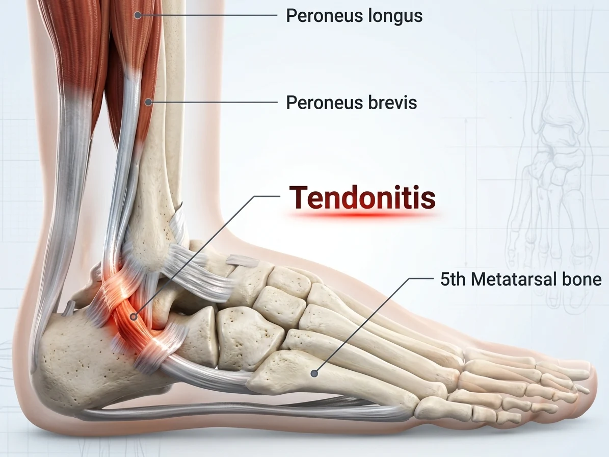
The terms peroneus longus and peroneus brevis are commonly used alongside the anatomical terms fibularis longus and fibularis brevis. All four terms may therefore appear in medical literature when discussing the peroneal muscles and their tendons.
Together, the peroneal tendons help evert the foot, meaning they turn the sole outward. They also contribute to dynamic ankle stability, particularly during walking, running, and activities performed on uneven ground.
What Is Peroneal Tendonitis?
Peroneal tendonitis refers to irritation and inflammation involving one or both of the peroneal tendons. It usually causes pain along the outer side of the ankle and may be associated with tenderness, swelling, or pain when the foot and ankle are moved or loaded.
The condition can be acute or chronic. Acute peroneal tendonitis develops over a relatively short period and may occur after an ankle sprain, excessive loading during sports, or prolonged walking on uneven surfaces. It may cause more noticeable pain, tenderness, and swelling.
Chronic peroneal tendonitis develops or persists over a longer period and may reflect ongoing tendon dysfunction and structural changes within the tendon rather than active inflammation alone.
The term peroneal tendinopathy is often preferred when describing a longer-lasting tendon disorder. Tendinopathy is a broader term that includes changes in the structure and function of the tendon and does not necessarily imply inflammation. For this reason, not every case of persistent peroneal tendon pain is caused by tendonitis in the strict inflammatory sense.
Peroneal tenosynovitis is a related but distinct condition. It refers to inflammation or irritation of the synovial sheath surrounding the tendon, rather than primarily affecting the tendon itself. Tendonitis and tenosynovitis can occur together, and their symptoms may overlap.
Although these terms describe somewhat different processes, they are often used interchangeably in everyday clinical practice and patient information. The underlying cause and factors that increase stress on the peroneal tendons are discussed in the next section.
What Causes Peroneal Tendonitis?
Peroneal tendonitis can develop when the peroneal tendons are exposed to more stress than they can tolerate and recover from. This may happen after a single ankle injury or gradually as a result of repetitive loading.
Ankle Sprains
An ankle sprain is a common trigger of peroneal tendon problems. When the ankle suddenly rolls, the peroneal tendons may be stretched forcefully as they work to stabilize the foot. In some cases, the tendons themselves may also be injured during the sprain.
Repetitive or Excessive Loading
Peroneal tendonitis can develop when the tendons are repeatedly exposed to loads that exceed their ability to recover. This may occur with running, jumping, rapid changes in direction, or prolonged walking and hiking on uneven surfaces.
The risk can increase when training volume or intensity is increased too quickly, when someone returns to sport after a period of inactivity, or when they suddenly take on a more demanding physical activity.
Anatomical Factors
Certain anatomical features around the ankle can increase the mechanical stress placed on the peroneal tendons. These include variations in the shape of the retromalleolar groove, a prominent peroneal tubercle, or accessory muscles and other structures that reduce the available space around the tendons.
Some people also have an os peroneum, a small accessory bone within the peroneus longus tendon. It is usually harmless, but in some cases it can contribute to irritation or tendon injury.
These anatomical variations do not necessarily cause symptoms on their own. Their clinical importance depends on how they affect tendon loading, movement, and the available space around the tendons.
Foot and Ankle Mechanics
The way the foot and ankle move during walking and running can influence the forces placed on the peroneal tendons. A hindfoot varus, in which the heel is tilted inward relative to the lower leg, can increase the demand on the peroneal muscles and tendons as they work to stabilize and evert the foot.
Other changes in foot and ankle mechanics may also alter the distribution of load across the peroneal tendons and contribute to persistent symptoms.
Previous Ankle Injuries
Previous ankle injuries can alter normal ankle movement and stability. Lateral ankle sprains, particularly injuries to the anterior talofibular ligament (ATFL), are a common cause of persistent ankle instability. If the ankle remains unstable after an injury, the peroneal tendons may have to work harder to provide dynamic support, potentially increasing their workload over time.
Other Peroneal Tendon Problems
Peroneal tendonitis may also occur alongside other tendon problems, including peroneal tendon tears or peroneal tendon subluxation. These conditions can cause similar symptoms and may sometimes develop together with tendon irritation.
For this reason, persistent or recurrent pain along the outer ankle should not automatically be attributed to tendonitis. A clinical examination and, when appropriate, imaging can help determine whether another peroneal tendon disorder is present.
Related Guides


Peroneal Tendonitis Symptoms
Typical symptoms of peroneal tendonitis include:
- Pain along the outer side of the ankle, often behind or just below the lateral malleolus
- Tenderness when pressing over the peroneal tendons
- Pain that increases with walking, running, or uneven-ground activities
- Swelling around the outer ankle
- Pain or discomfort when turning the foot outward
- A feeling of weakness or instability around the ankle
- In some cases, snapping or clicking behind the outer ankle, particularly when the tendons sublux or dislocate
How Is Peroneal Tendonitis Diagnosed?
Peroneal tendonitis is usually diagnosed based on the symptoms, medical history, and physical examination. The clinician will assess the location of pain and tenderness, ankle stability, and how the peroneal tendons respond to movement and loading.
Common parts of the evaluation include:
- Medical history – previous ankle sprains, recent changes in activity, and the onset and pattern of symptoms
- Physical examination – tenderness, swelling, ankle range of motion, and pain during resisted foot eversion
- Ankle stability testing – particularly after previous lateral ankle sprains or suspected ATFL injury
- Ultrasound – can show tendon thickening, changes in tendon structure, fluid around the tendon sheath, and tendon tears or subluxation
- MRI – may be used when the diagnosis is uncertain or when a tendon tear, significant structural abnormality, or another cause of lateral ankle pain is suspected
X-rays do not show the peroneal tendons directly but may be useful when a bone injury, fracture, or anatomical abnormality is suspected.
How Is Peroneal Tendonitis Treated?
Treatment depends on the severity of the tendon problem, how long symptoms have been present, and whether there is an associated tendon tear or ankle instability. Most cases are initially treated without surgery.
Activity Modification
The first step is to reduce activities that aggravate the tendon, particularly running, jumping, and prolonged walking on uneven ground. Complete rest is usually unnecessary. As symptoms improve, activity should be gradually increased.
Physical Therapy
Physical therapy is the mainstay of treatment. Rehabilitation focuses on gradually loading the peroneal tendons, strengthening the ankle and lower leg, and improving balance and proprioception. The program should progress according to pain and function rather than relying on prolonged rest.
Bracing and Immobilization
An ankle brace can reduce painful loading and provide additional stability, particularly when ankle instability is present. Short-term immobilization in a walking boot may be considered for more painful acute cases or associated tendon injury, but prolonged immobilization is generally avoided in uncomplicated tendinopathy.
Medications
NSAIDs may be useful for short-term pain relief, particularly when symptoms are accompanied by acute inflammation or swelling. They should not be considered a treatment that repairs the underlying tendon problem.
Injections
Injections are not routinely required. An ultrasound-guided corticosteroid injection into the peroneal tendon sheath may be considered in selected patients with persistent pain or tenosynovitis, but evidence is limited and potential tendon-related risks should be considered. PRP is not currently supported by sufficient evidence for routine treatment of peroneal tendinopathy.
Surgery
Surgery is generally reserved for patients who do not improve with appropriate conservative treatment or who have a significant peroneal tendon tear, rupture, or tendon instability. Depending on the underlying problem, surgery may involve tendon repair or debridement, stabilization of the tendons, or correction of an anatomical abnormality contributing to the disorder.
How Long Does Peroneal Tendonitis Take to Heal?
Recovery time varies depending on the severity of the tendon problem, its duration, and whether there is an associated tendon tear or ankle instability.
Mild cases may improve within several weeks with appropriate activity modification and rehabilitation. More persistent or severe cases can take several months to recover, particularly when tendon damage or chronic ankle instability is present.
A gradual return to normal activities is generally preferable to returning to full activity as soon as the pain subsides.
When Should You See a Doctor?
Consider seeing a doctor if:
- Pain persists despite reducing aggravating activities
- Symptoms are getting worse rather than improving
- There is significant swelling or bruising around the ankle
- You cannot walk normally or put weight on the foot
- The ankle repeatedly gives way
- You notice snapping or a feeling that the tendon moves out of place
- Symptoms began after a significant ankle injury
Prompt medical assessment is particularly important when a peroneal tendon tear, tendon subluxation, or significant ankle ligament injury is suspected.
Frequently Asked Questions
Can you walk with peroneal tendonitis?
Yes. Many people with peroneal tendonitis can walk, although walking may be painful, particularly on uneven surfaces or during longer periods of activity. Severe symptoms may temporarily require reduced weight-bearing or immobilization.
What does peroneal tendonitis feel like?
It typically causes pain and tenderness along the outer side of the ankle, often behind or below the lateral malleolus. Pain may increase with walking, running, or movements that load the peroneal tendons.
Can peroneal tendonitis heal on its own?
Mild cases may improve with appropriate activity modification and gradual rehabilitation. Persistent symptoms, recurrent ankle instability, or suspected tendon damage should be evaluated by a healthcare professional.
Is walking good for peroneal tendonitis?
Walking can usually be continued as tolerated if it does not significantly increase symptoms. However, prolonged walking, running, and uneven-ground activities may need to be reduced temporarily while the tendon recovers.
Can peroneal tendonitis cause ankle instability?
Peroneal tendon problems can contribute to ankle instability, but instability is also commonly related to previous lateral ankle ligament injuries, particularly ATFL injury. When both conditions are present, treatment may need to address the tendon and the underlying ankle instability.
van Dijk PA et al. The ESSKA-AFAS international consensus statement on peroneal tendon pathologies. Knee Surgery, Sports Traumatology, Arthroscopy. Clinical consensus: Diagnosis and treatment of peroneal tendon disorders
van Dijk PA et al. Peroneal tendon disorders. EFORT Open Reviews. Clinical review: Peroneal tendon anatomy, diagnosis and treatment
Danna NR, Brodsky JW. Diagnosis and Operative Treatment of Peroneal Tendon Tears. Foot & Ankle Orthopaedics. Clinical review: Peroneal tendon tears, imaging and surgical treatment
Chhabra A et al. Peroneal tendon pathology: Pre- and post-operative high resolution US and MR imaging. European Journal of Radiology. Imaging review: Ultrasound and MRI of peroneal tendon pathology
Major NM et al. Ultrasound and MRI of the peroneal tendons and associated pathology. Seminars in Musculoskeletal Radiology. Imaging guide: Ultrasound and MRI evaluation of the peroneal tendons
Stewart CM et al. Diagnostic accuracy of MRI and US for peroneal tendon tears: a systematic review and meta-analysis. Systematic review: Diagnostic accuracy of ultrasound and MRI for peroneal tendon tears