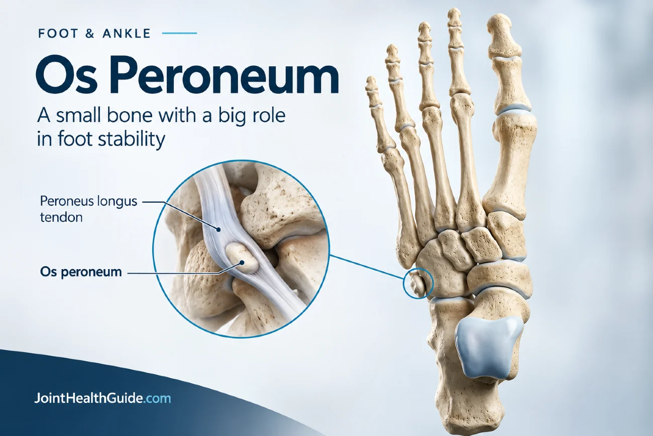
Os peroneum is a small accessory bone located within the peroneus longus tendon on the outer side of the foot, close to the cuboid bone. It is a relatively common anatomical variant and, in most people, causes no symptoms and requires no treatment. It is often discovered incidentally on an X-ray or other imaging study performed for an unrelated foot or ankle problem.
However, the os peroneum can become clinically important when it is injured or when it is associated with damage to the peroneus longus tendon. A fracture, displacement, or chronic stress involving the ossicle may cause pain on the outer side of the midfoot. In some cases, an abnormal os peroneum can also be a clue to a partial or complete tear of the peroneus longus tendon.
Painful os peroneum syndrome is a term used for these symptomatic conditions. Because lateral foot pain has many possible causes, recognizing the characteristic location of the os peroneum and its relationship to the peroneus longus tendon can help guide the diagnosis and treatment.
What Is the Os Peroneum?
The os peroneum is a small accessory bone located within the peroneus longus tendon, on the outer side of the foot near the cuboid bone. It is classified as an accessory ossicle and is a normal anatomical variant rather than a disease.
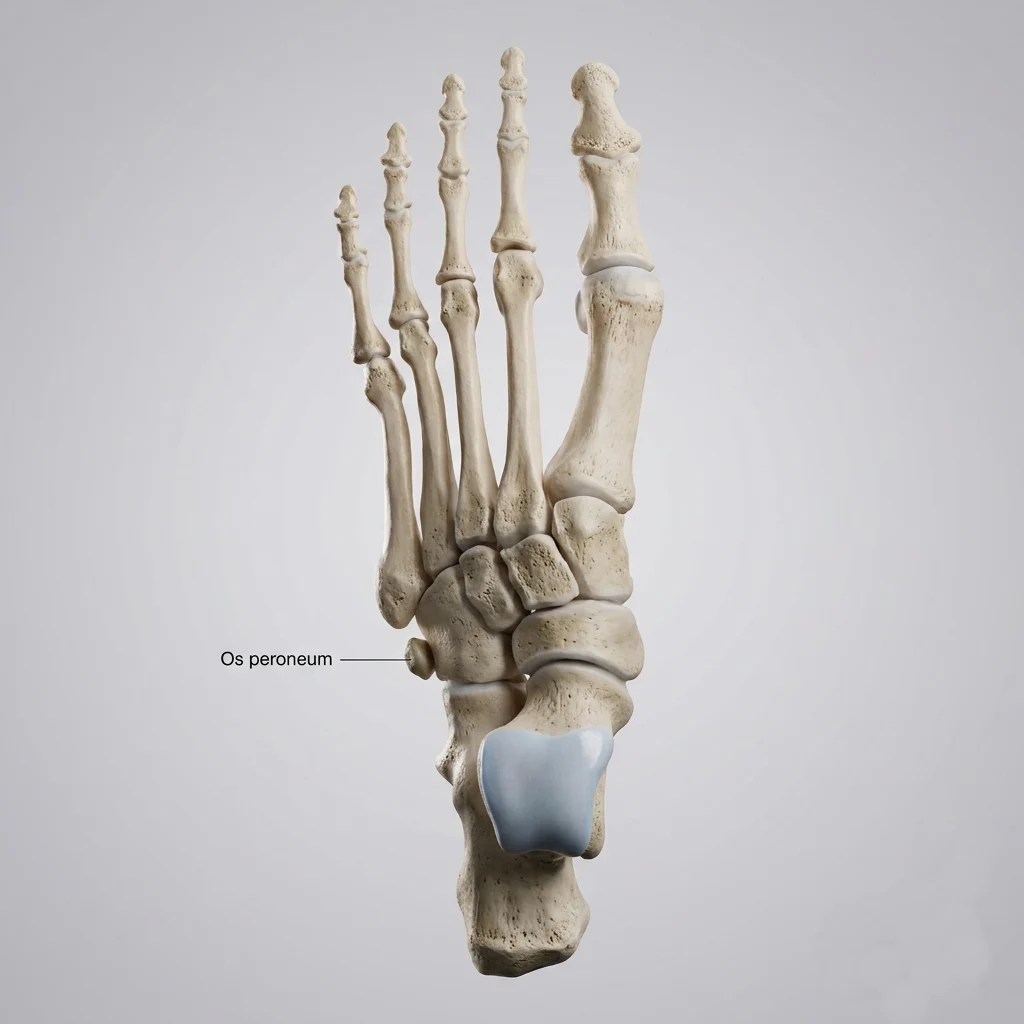
The os peroneum is found in approximately 5% to 30% of people and is often present in both feet. Most cases are asymptomatic and discovered incidentally on an X-ray performed for another foot or ankle problem.
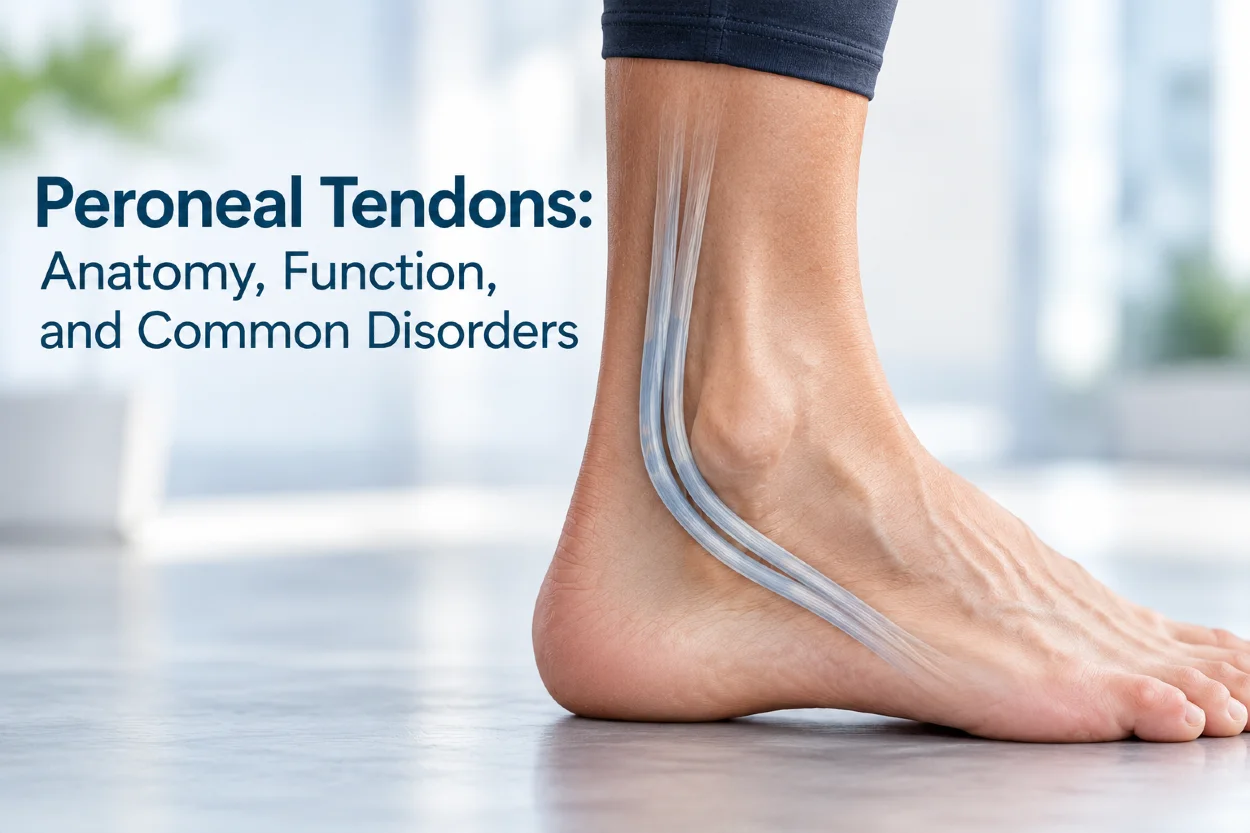
The peroneus longus tendon runs behind the outer ankle, along the lateral foot, and underneath the cuboid toward its attachment at the base of the first metatarsal and medial cuneiform. It helps with foot eversion, plantarflexion, and foot stability.
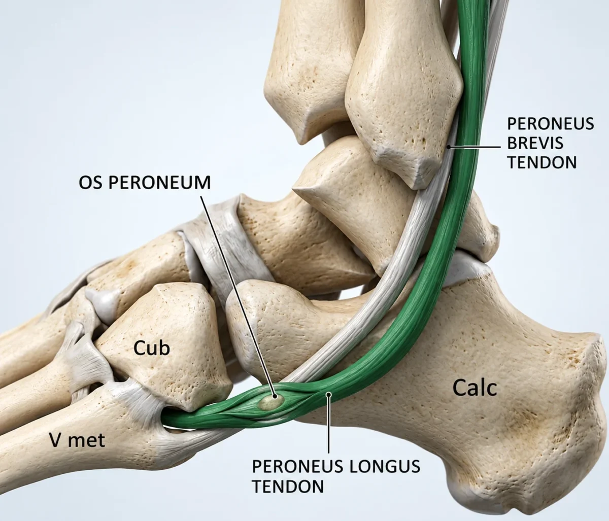
Because the os peroneum lies within this tendon, a fracture or displacement of the bone may indicate an associated peroneus longus tendon injury. However, its presence on an X-ray does not necessarily explain foot pain. Symptoms, imaging findings, and the condition of the surrounding tendon must be assessed together.
Is Os Peroneum Always a Problem?
No. In most people, an os peroneum is a normal, painless anatomical variant and does not require treatment. Its presence on an X-ray does not necessarily mean that it is responsible for foot pain.
Problems arise when the os peroneum is injured or becomes involved with pathology of the peroneus longus tendon. This can occur after an acute ankle or foot injury, repetitive overuse, or chronic tendon degeneration.
An os peroneum may become fractured, displaced, or associated with peroneus longus tendinopathy, tenosynovitis, or a tendon tear. These conditions can cause pain on the outer side of the midfoot, particularly during walking, running, or activities that load the peroneal tendons.
Clinical Insight
Therefore, finding an os peroneum on imaging is only one part of the diagnosis. Its shape, position, and relationship to the peroneus longus tendon should be considered together with the patient’s symptoms and physical examination.
What Is Painful Os Peroneum Syndrome?
Painful os peroneum syndrome (POPS) is a term used to describe lateral midfoot pain associated with an abnormal or injured os peroneum and/or the surrounding peroneus longus tendon. It is not a single disease but a clinical syndrome with several possible causes.
Pain may develop after an acute injury, such as an ankle sprain or sudden forceful contraction of the peroneus longus tendon. Repetitive loading can also cause stress-related injury or inflammation over time. The os peroneum may become fractured, displaced, or associated with peroneus longus tendinopathy, tenosynovitis, or a partial or complete tendon tear.
Symptoms typically include pain and tenderness along the outer aspect of the midfoot, near the cuboid. Swelling may occur, and pain can increase with walking, running, or activities that load the peroneal tendons.
Importantly, the presence of an os peroneum alone does not mean that POPS is present. The diagnosis depends on the clinical picture and imaging findings, particularly whether there is evidence of injury to the ossicle or the peroneus longus tendon.
Causes and Risk Factors
Problems involving the os peroneum usually develop because of injury or overload of the peroneus longus tendon rather than simply because the accessory bone is present.
An acute injury can occur when the foot is forced into inversion and supination, particularly during a sudden contraction of the peroneus longus tendon. This mechanism can fracture the os peroneum and, in more severe cases, cause disruption of the tendon.
Repetitive loading can also contribute to chronic irritation and degeneration. Activities involving frequent running, jumping, or changes in direction may place increased stress on the peroneal tendons. Over time, this may lead to tendinopathy, tenosynovitis, or a tendon tear involving the region around the os peroneum.
Several anatomical and mechanical factors may increase the risk of peroneal tendon problems:
- Cavovarus foot alignment, particularly a varus hindfoot, which can increase stress on the lateral structures of the foot.
- Previous ankle injuries, which may alter foot mechanics and increase chronic tendon stress.
- Previous peroneal tendon injury, which can predispose the tendon to further damage.
- Repetitive sports activity, especially activities involving running, jumping, or frequent changes in direction.
- Age-related tendon degeneration, which may reduce the tendon’s ability to tolerate repetitive loading.
Importantly, the presence of an os peroneum itself is not a disease or an independent reason for symptoms.
Symptoms
Symptoms vary depending on whether the os peroneum itself is injured or whether there is associated peroneus longus tendon pathology. Common symptoms include:
- Pain on the outer side of the midfoot, typically near the cuboid and along the course of the peroneus longus tendon.
- Tenderness over the os peroneum or surrounding tendon.
- Swelling around the lateral midfoot, particularly after an acute injury.
- Pain with walking, running, or push-off, especially when the peroneal tendons are loaded.
- Weakness with foot eversion or plantarflexion, which may suggest more significant peroneus longus tendon involvement.
- A snapping or instability sensation may occur when there is associated peroneal tendon subluxation.
The severity of symptoms does not necessarily reflect the size or appearance of the os peroneum. The underlying tendon injury is often an important determinant of symptoms and functional limitation.
Os Peroneum Fracture
An os peroneum fracture is an uncommon but important cause of lateral midfoot pain. It typically occurs when the peroneus longus tendon contracts forcefully while the foot is suddenly forced into inversion and supination.
A fracture may occur in isolation, but it can also be associated with a partial or complete peroneus longus tendon tear. This is clinically important because displacement of the os peroneum can provide a clue to the severity of the underlying tendon injury.
On X-ray, an acute fracture may appear as an irregular or sharply defined break through the normally smooth, well-corticated ossicle. Separation of fragments greater than approximately 6 mm raises suspicion for an associated tendon tear, although this finding should always be interpreted in the context of the clinical examination and other imaging findings.
Not every fragmented os peroneum represents an acute fracture. A bipartite or multipartite os peroneum may have smooth, well-corticated margins and can resemble a fracture on imaging. Comparison with the opposite foot and correlation with the mechanism of injury can help distinguish these conditions.
Diagnosis
Diagnosing a symptomatic os peroneum requires more than simply identifying the ossicle on an X-ray. The main question is whether the os peroneum or the peroneus longus tendon is responsible for the patient’s lateral foot pain.
The evaluation begins with the patient’s history and physical examination. The clinician should consider the mechanism of injury, particularly a sudden inversion or supination injury, as well as repetitive sports activity or a previous ankle injury. Tenderness is usually located along the peroneus longus tendon near the cuboid. Foot alignment, peroneal muscle strength, tendon stability, and signs of associated ankle or foot injuries should also be assessed.
Because several conditions can cause pain in this region, the differential diagnosis may include:
- Peroneal tendinopathy or tendon tear
- Peroneal tendon subluxation
- Lateral ankle ligament injury
- Cuboid syndrome
- Cuboid or fifth metatarsal fracture
- Other accessory ossicles that can mimic an os peroneum
Plain X-rays are usually the first imaging test. The os peroneum typically appears as a small, round or oval, well-corticated ossicle near the cuboid and is often best seen on an oblique view. An acute fracture may produce an irregular or sharply defined fracture line. Fragment separation of more than approximately 6 mm raises suspicion for an associated peroneus longus tendon tear, while proximal migration of the ossicle may also indicate tendon disruption.
However, a fragmented appearance does not automatically mean that an acute fracture is present. A bipartite or multipartite os peroneum may have smooth, well-corticated margins and can resemble a fracture. Comparison with the opposite foot can be helpful when the diagnosis is uncertain.
Musculoskeletal ultrasound can provide detailed and dynamic assessment of both the os peroneum and the peroneus longus tendon. The ossicle appears as a hyperechoic structure with posterior acoustic shadowing. Ultrasound can demonstrate tendon thickening, tenosynovitis, partial or complete tears, tendon retraction, and displacement of the os peroneum. Dynamic scanning can also identify peroneal tendon subluxation. Comparing the symptomatic side with the opposite foot may provide additional information.
MRI is useful when more detailed assessment of the tendon and surrounding soft tissues is required. It can demonstrate peroneus longus tendinopathy, tenosynovitis, tendon tears, and bone marrow edema within an injured os peroneum. CT provides excellent visualization of the bony anatomy and cortical margins and can help distinguish a fracture from a bipartite or multipartite ossicle.
Clinical Insight
Ultimately, the diagnosis depends on correlating the imaging findings with the patient’s symptoms, physical examination, and mechanism of injury. The most important finding is often not simply the presence of an os peroneum, but evidence of associated peroneus longus tendon pathology.
Treatment
Treatment depends on the specific pathology and the severity of the symptoms. An asymptomatic os peroneum requires no treatment. When pain is caused by irritation, tendinopathy, or a minor injury, conservative treatment is usually the first approach.
- Tendinopathy or tenosynovitis: Activity modification, temporary reduction of tendon loading, pain control, and progressive physical therapy are usually appropriate. Foot biomechanics and cavovarus alignment should also be addressed when relevant.
- Minor or nondisplaced os peroneum injury: A period of relative rest or temporary immobilization may be used, followed by gradual rehabilitation as symptoms improve.
- Peroneus longus tendon tear: Partial tears may sometimes be managed conservatively, depending on their severity and functional impact. Significant or complete tears may require surgical tendon repair or reconstruction.
- Displaced os peroneum or fracture with tendon disruption: Significant displacement, persistent pain, or associated tendon injury may require surgery. Depending on the injury, treatment may include excision of the os peroneum with tendon repair or tenodesis, or fixation in selected fractures.
- Persistent symptoms despite conservative treatment: Surgical treatment may be considered when pain and functional limitation continue despite appropriate nonoperative management.
The goal is not simply to remove the os peroneum, but to treat the underlying source of pain and restore the function of the peroneus longus tendon. The appropriate treatment therefore depends on the integrity of the tendon, the position and condition of the os peroneum, and the patient’s functional demands.
Prognosis and When to See a Doctor
The prognosis of an os peroneum problem depends mainly on the underlying peroneus longus tendon injury and the severity of the associated bone or soft-tissue damage. Minor injuries and tendon irritation often improve with appropriate activity modification and rehabilitation, while significant tendon tears or displaced fractures may require a longer recovery and, in some cases, surgery.
Because lateral foot pain has many possible causes, persistent or worsening symptoms should not simply be attributed to an incidental os peroneum found on an X-ray.
Medical evaluation is particularly appropriate when:
- Pain persists after an ankle or foot injury.
- There is significant swelling, bruising, or difficulty bearing weight.
- Pain is localized over the course of the peroneus longus tendon.
- There is weakness with foot eversion or plantarflexion.
- Symptoms include snapping or a sensation of tendon instability.
- Pain continues despite rest and initial conservative treatment.
Early assessment can help identify a peroneus longus tendon tear or other significant injury and prevent prolonged symptoms or functional limitation.
Key Clinical Points – Os Peroneum
- Os peroneum is a common accessory bone located within the peroneus longus tendon near the cuboid.
- In most people, the os peroneum is a normal anatomical variant and does not cause symptoms or require treatment.
- Painful os peroneum syndrome occurs when the ossicle or the surrounding peroneus longus tendon becomes injured, inflamed, or damaged.
- An abnormal or displaced os peroneum can be an important clue to a peroneus longus tendon tear, particularly after an acute injury.
- X-ray, ultrasound, MRI, and CT can help distinguish an os peroneum fracture from a normal or multipartite ossicle and identify associated tendon pathology.
- Treatment depends on the underlying problem and may range from activity modification and rehabilitation to surgical treatment for significant tendon tears, displaced fractures, or persistent symptoms.
Frequently Asked Questions
Is an os peroneum a bone?
Yes. The os peroneum is a small accessory bone located within the peroneus longus tendon near the cuboid. It is a normal anatomical variant and is not a disease.
Can an os peroneum cause foot pain?
Yes, but the presence of an os peroneum alone does not usually cause pain. Pain may occur when the ossicle is fractured, displaced, or associated with peroneus longus tendon injury.
What does a fractured os peroneum feel like?
A fractured os peroneum can cause pain and tenderness on the outer side of the midfoot, sometimes accompanied by swelling and difficulty walking. Pain may increase when the peroneal tendons are loaded.
How is an os peroneum fracture diagnosed?
X-rays are usually the first imaging test. Ultrasound, MRI, or CT may be used when the diagnosis is uncertain or when an associated peroneus longus tendon tear needs to be assessed.
Does a fractured os peroneum need surgery?
Not always. Minor or nondisplaced injuries may improve with activity modification, immobilization, and rehabilitation. Surgery may be considered for significant displacement, tendon disruption, persistent pain, or major functional impairment.
How long does it take to recover from an os peroneum injury?
Recovery varies depending on the severity of the injury and whether the peroneus longus tendon is damaged. Minor injuries may improve with conservative treatment, while tendon tears or surgical cases generally require a longer rehabilitation period.
Hindi HF, Byerly DW. Os Peroneum. StatPearls [Internet]. Treasure Island (FL): StatPearls Publishing; 2023 Jan-. Updated February 5, 2023. NCBI Bookshelf: Os Peroneum – diagnosis, imaging, and treatment
Bianchi S, Bortolotto C, Draghi F. Os peroneum imaging: normal appearance and pathological findings. Insights Imaging. 2017;8:59-68. PubMed: Os peroneum imaging and pathological findings
Sobel M, Pavlov H, Geppert MJ, Thompson FM, DiCarlo EF, Davis WH. Painful os peroneum syndrome: a spectrum of conditions responsible for plantar lateral foot pain. Foot Ankle Int. 1994;15(3):112-124. PubMed: Painful os peroneum syndrome and lateral foot pain
The ESSKA-AFAS International Consensus Group. The ESSKA-AFAS international consensus statement on peroneal tendon pathologies. PMC: Consensus recommendations for peroneal tendon disorders and painful os peroneum syndrome
Segatt PH, Prado JLMDA, Duarte ML. Painful Os Peroneum Syndrome: An Unusual Diagnosis. Kurume Med J. 2025;71(3.4):153-156. doi:10.2739/kurumemedj.MS7134006. PubMed: Painful Os Peroneum Syndrome – 2025 case report