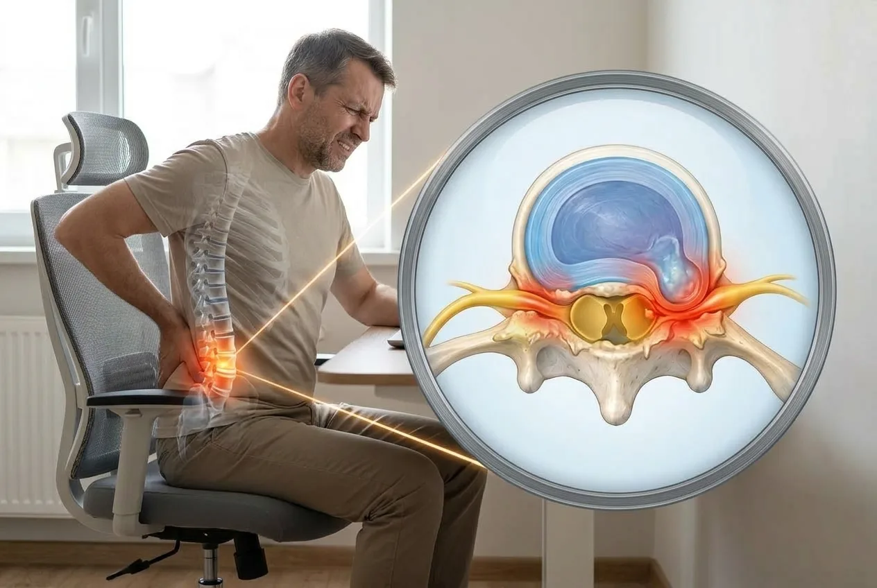
Foraminal stenosis is a condition in which one or more openings between the vertebrae become narrowed, potentially compressing the spinal nerves that pass through them. These openings, called neural foramina, are located on both sides of the spine and provide a passageway for nerve roots as they leave the spinal cord and travel to the rest of the body. Foraminal stenosis most commonly affects the cervical spine (neck) or lumbar spine (lower back).
Narrowing of the neural foramen does not always cause symptoms. However, when the space becomes too small and irritates or compresses a nerve root, it can lead to pain, tingling, numbness, or weakness in the arm or leg. The symptoms depend largely on which spinal nerve is affected and whether the narrowing occurs in the neck or lower back.
Foraminal stenosis is sometimes confused with spinal canal stenosis, but these conditions are not identical. Foraminal stenosis involves narrowing of the opening where an individual spinal nerve exits the vertebral column, whereas spinal canal stenosis refers to narrowing of the central spinal canal that contains the spinal cord and nerve roots. Understanding this distinction is important because the symptoms and treatment considerations can differ between the two conditions.
Quick Overview – Foraminal Stenosis
- Foraminal stenosis is narrowing of the openings through which spinal nerves exit the spine.
- The condition most commonly affects the cervical spine (neck) and lumbar spine (lower back), although it can occur at other spinal levels.
- Common causes include age-related degeneration, disc degeneration, bone spurs, facet joint arthritis, and herniated discs.
- Many people have foraminal narrowing without symptoms. Symptoms develop when the narrowed foramen irritates or compresses a spinal nerve.
- Symptoms may include radiating pain, tingling, numbness, or muscle weakness in the arm or leg, depending on the affected nerve root.
- MRI is the preferred imaging test, but imaging findings must be interpreted together with symptoms and neurological examination.
- Treatment often begins with conservative measures, including physical therapy, activity modification, and medications when appropriate. Injections or surgery may be considered in selected cases.
- Severe foraminal stenosis on MRI does not automatically require surgery. Treatment decisions depend on symptoms, neurological findings, and functional limitations.
What Is Foraminal Stenosis?
Foraminal stenosis is the narrowing of the openings in the spine through which spinal nerves leave the spinal canal. These openings are called neural foramina (singular: neural foramen). When one of these spaces becomes narrower, the resulting condition is known as neural foraminal stenosis or foraminal narrowing.
In many people, foraminal narrowing develops gradually as part of the normal aging process and does not cause any symptoms. However, if the narrowing reduces the available space around a spinal nerve root, it can cause irritation or compression of the nerve. This may result in radiating pain, tingling, numbness, or muscle weakness in the area supplied by that nerve.
The location of foraminal stenosis determines which symptoms may develop. Cervical foraminal stenosis affects the neck region and can cause symptoms in the shoulder, arm, or hand. Lumbar foraminal stenosis affects the lower back and may cause pain, numbness, or weakness extending into the buttock or leg.
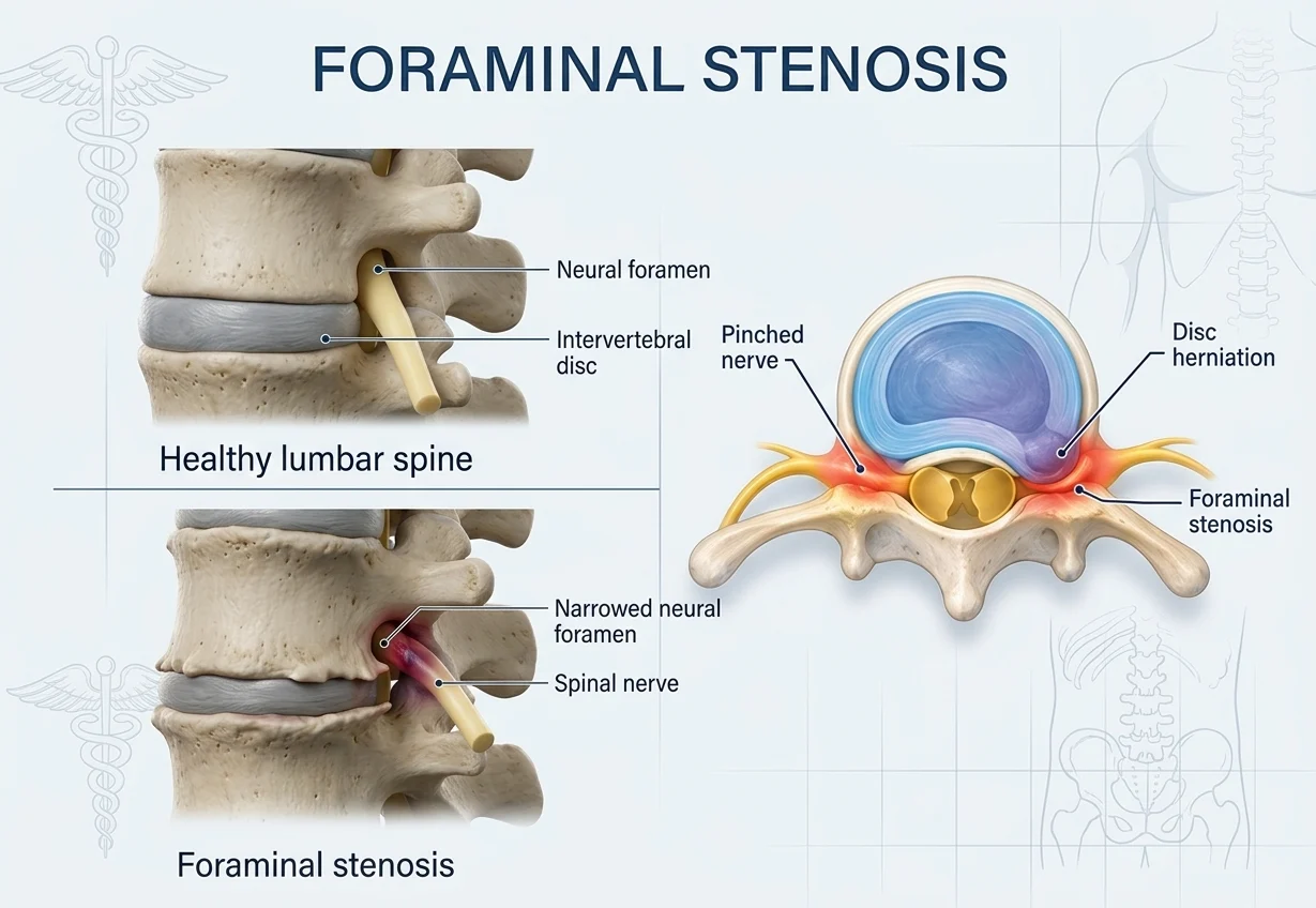
What Is the Neural Foramen?
The neural foramen is an opening between adjacent vertebrae through which a spinal nerve root exits the spinal canal. There is one foramen on each side of the vertebral column at most spinal levels.
The term neural foramen refers to a single opening, while neural foramina is the plural form. When degenerative changes such as bone spurs or disc degeneration narrow these openings, the resulting condition is called foraminal stenosis.
What Causes Foraminal Stenosis?
Foraminal stenosis usually develops because of structural changes in the spine that gradually reduce the space available for a spinal nerve root. Although aging is the most common underlying factor, several other conditions can contribute to neural foraminal narrowing.
Age-Related Degenerative Changes
Degenerative changes, also called spondylosis, associated with aging are the most common cause of foraminal stenosis. Over time, the intervertebral discs lose height and elasticity, while the facet joints and surrounding bones may develop arthritis.
These changes can narrow the neural foramen through several mechanisms:
- Degenerative disc disease: Loss of disc height can reduce the vertical space within the foramen.
- Bone spurs (osteophytes): Extra bone growth along the vertebral bodies or facet joints can protrude into the neural foramen.
- Facet joint arthritis: Enlargement and degeneration of the facet joints may reduce the available space for the exiting nerve root.
- Ligament thickening: Degenerative changes can cause spinal ligaments to become thicker and less flexible, potentially contributing to narrowing of the neural foramen. The ligamentum flavum is more commonly implicated in central spinal canal stenosis.
These processes often occur together, particularly in the cervical and lumbar spine.
Herniated or Bulging Disc
A herniated or bulging intervertebral disc can extend toward the neural foramen and reduce the space available for a spinal nerve. Depending on its location, a disc herniation may cause foraminal stenosis, lateral recess stenosis, or central spinal canal stenosis.
Disc-related narrowing may develop suddenly or gradually and is a common cause of nerve root compression in both the neck and lower back.
Spondylolisthesis
Spondylolisthesis occurs when one vertebra slips forward or backward relative to an adjacent vertebra. This altered alignment can change the dimensions of the neural foramen and reduce the space available for the exiting nerve root.
Less Common Causes
Foraminal stenosis can sometimes result from less common conditions, including congenital or developmental narrowing of the neural foramina. In these cases, the available space around a nerve may already be limited, making the nerve more susceptible to compression when degenerative changes develop later in life.
Other less common causes include spinal fractures, tumors, infections, and inflammatory conditions affecting the spine. These conditions require specific evaluation because treatment depends on the underlying cause.
Related Guides


Cervical Foraminal Stenosis
Cervical foraminal stenosis occurs when one of the openings in the neck becomes narrowed and reduces the space available for a spinal nerve. The narrowing may develop gradually as the discs, joints, and bones in the cervical spine change with age.
When the narrowed space irritates or compresses a nerve, symptoms may extend beyond the neck. Instead of feeling pain only in the neck, you may notice pain traveling into the shoulder, arm, or hand. Depending on which nerve is affected, you may also experience tingling, numbness, or weakness in specific parts of the arm or hand.
What Does Cervical Foraminal Stenosis Feel Like?
Symptoms can vary considerably from person to person. Common symptoms include:
- Neck pain, sometimes with stiffness
- Pain that travels into the shoulder or arm
- Tingling or pins and needles in the arm or hand
- Numbness in part of the arm or hand
- Muscle weakness, such as reduced grip strength or difficulty lifting the arm
- Symptoms that become worse with certain neck movements in some people
The exact location of the symptoms depends on which nerve is affected. For example, compression of a nerve in the lower part of the cervical spine can cause symptoms that extend farther down the arm and into the hand.
When cervical foraminal stenosis irritates a spinal nerve and produces radiating pain, numbness, tingling, or weakness, it can lead to cervical radiculopathy. Learn more about cervical radiculopathy.
Importantly, cervical stenosis seen on an MRI does not necessarily mean that you will have symptoms. Some people have narrowing of the neural foramina without significant nerve compression or related complaints.
Lumbar Foraminal Stenosis
Lumbar foraminal stenosis occurs when the opening through which a spinal nerve leaves the lower back becomes narrowed. As in the neck, this narrowing may develop gradually because of age-related changes in the discs, joints, and bones of the spine.
When a lumbar nerve is irritated or compressed, symptoms may extend from the lower back into the buttock and leg. Lumbar foraminal stenosis can therefore cause sciatica or sciatica-like symptoms, particularly when it affects a nerve root that contributes to the sciatic nerve. Some people mainly notice pain, while others experience numbness, tingling, or weakness. The pattern depends on which nerve is affected.
What Does Lumbar Foraminal Stenosis Feel Like?
Common symptoms include:
- Low back pain
- Pain traveling into the buttock or leg, sometimes resembling sciatica
- Tingling or numbness in the leg or foot
- Weakness in the leg or foot
- Symptoms that may become worse with certain positions or movements
Not everyone with lumbar foraminal narrowing has symptoms. Mild or even severe narrowing seen on an MRI may sometimes be present without significant nerve compression or related complaints.
Foraminal Stenosis vs. Lateral Recess Stenosis
Both conditions can compress a spinal nerve, but they occur in different parts of the spine.
Foraminal stenosis affects the opening where the nerve leaves the spinal canal. Lateral recess stenosis affects the passageway inside the spinal canal through which the nerve travels before reaching the neural foramen.
This distinction matters because the location of the narrowing can help explain which part of the nerve is affected and where symptoms develop.
If you want to learn more about spinal stenosis, including its causes, symptoms, diagnosis, and treatment options, read our guide to spinal stenosis.
Foraminal Stenosis Symptoms
Foraminal stenosis does not always cause symptoms. Symptoms develop when narrowing of the neural foramen irritates or compresses a spinal nerve. Because each nerve supplies a different area of the body, the symptoms depend on the level of the spine that is affected.
| Spinal Region | Possible Symptoms |
|---|---|
| Cervical spine (neck) | Neck pain, shoulder or arm pain, tingling, numbness, or weakness in the arm or hand |
| Thoracic spine (mid-back) | Pain, tingling, or numbness that may wrap around the chest or abdomen; symptoms are less common |
| Lumbar spine (lower back) | Low back pain, buttock or leg pain, tingling, numbness, or weakness in the leg or foot |
The symptoms may occur on one side or, less commonly, on both sides, depending on whether one or multiple nerve roots are affected.
Does Foraminal Stenosis Always Cause Symptoms?
No. Foraminal narrowing can be visible on an MRI or CT scan without causing noticeable symptoms. Degenerative changes are common with aging, and imaging findings do not always correspond to the severity of a person’s pain or neurological symptoms.
For this reason, doctors interpret imaging findings together with the patient’s symptoms and physical examination rather than diagnosing symptomatic foraminal stenosis based on a scan alone.
Related Guides


Severe Foraminal Stenosis: What Does It Mean?
MRI reports may describe foraminal stenosis as mild, moderate, or severe, referring to the degree of narrowing and the amount of space remaining around the exiting spinal nerve. The exact grading can vary between radiologists.
Severe foraminal stenosis means that the neural foramen is substantially narrowed, potentially leaving little space around the nerve. However, the severity seen on an MRI does not always correspond to the severity of symptoms. Some people with severe narrowing have few or no symptoms, while others may experience significant nerve-related pain, numbness, or weakness.
Clinical Insight
Therefore, severe foraminal stenosis on an MRI does not automatically mean that surgery is required. Treatment depends on the symptoms, neurological findings, functional limitations, and response to conservative treatment.
How Is Foraminal Stenosis Diagnosed?
Diagnosis starts with a medical history and physical examination. Your doctor will ask about the location and pattern of your pain and look for sensory changes, muscle weakness, and changes in reflexes. Certain movements or physical tests may also help determine whether a spinal nerve is being irritated.
MRI is the main imaging test for evaluating foraminal stenosis because it can show the neural foramina, spinal nerves, intervertebral discs, and surrounding soft tissues. It can help identify whether a narrowed foramen is associated with nerve root compression and show degenerative changes such as disc degeneration or bone spurs.
CT provides more detailed images of bone and can be particularly useful when bony changes are contributing to foraminal narrowing. X-rays can show alignment, loss of disc height, and other degenerative changes, but they cannot directly visualize spinal nerves or reliably determine whether a nerve is being compressed.
Clinical Insight
Foraminal stenosis may be suspected clinically based on the pattern of pain, sensory changes, weakness, and other signs of nerve irritation. However, the narrowing itself can only be confirmed with imaging. MRI is the preferred imaging method because it can visualize the neural foramina, nerve roots, discs, and other soft tissues. CT can be used when MRI is contraindicated or when more detailed assessment of the bony structures is needed.
Treatment for Foraminal Stenosis
Treatment depends on the severity of the narrowing, the symptoms it causes, and whether there is evidence of nerve dysfunction. Many people with symptomatic foraminal stenosis can initially be treated without surgery.
Conservative Treatment
Conservative treatment may include temporarily modifying activities that aggravate symptoms, physical therapy, and targeted therapeutic exercises. Treatment may focus on improving strength, mobility, posture, and movement patterns while avoiding positions that significantly increase nerve irritation.
Pain-relieving or anti-inflammatory medications such as NSAIDs may also be used when appropriate, taking into account individual contraindications and risks.
Injections
When pain remains significant despite initial treatment, spinal injections may sometimes be considered. Depending on the location and clinical situation, options include epidural steroid injections and selective nerve root blocks.
A transforaminal injection delivers medication close to the affected nerve root and may also have a diagnostic role in selected patients.
Surgery
Surgery may be considered when symptoms remain severe and disabling despite appropriate conservative treatment, or when there is progressive neurological deficit. The specific procedure depends on the location and cause of the nerve compression and may involve removing the structure compressing the nerve and, when necessary, stabilizing the affected spinal segment.
Importantly, the presence of foraminal stenosis on imaging alone is not an indication for surgery. Symptoms, neurological findings, and imaging results need to be considered together when deciding on treatment.
Foraminal Stenosis vs. Spinal Canal Stenosis
Although both conditions involve narrowing within the spine, they affect different spaces and can produce different patterns of symptoms. Foraminal stenosis narrows the opening through which an individual spinal nerve exits the spine, while spinal canal stenosis narrows the central canal.
| Feature | Foraminal Stenosis | Spinal Canal Stenosis |
|---|---|---|
| What is narrowed? | The opening where a spinal nerve exits the spine | The central spinal canal |
| Typical nerve involvement | Usually an individual exiting nerve root | The spinal cord or multiple nerve roots, depending on the location |
| Common symptoms | Pain, tingling, numbness, or weakness along the affected nerve | Symptoms vary by location and may include neurogenic claudication or, in the cervical spine, symptoms related to spinal cord compression |
The two conditions can also occur at the same time. For example, degenerative changes in the lumbar spine may narrow both the central spinal canal and the neural foramina. This is why imaging findings need to be interpreted together with the patient’s symptoms and neurological examination.
Frequently Asked Questions About Foraminal Stenosis
What is foraminal stenosis?
Foraminal stenosis is narrowing of the opening between the vertebrae through which a spinal nerve exits the spine. If the narrowing compresses or irritates the nerve, it can cause pain, tingling, numbness, or weakness.
Is foraminal stenosis serious?
Foraminal stenosis is not always serious. Some people have significant narrowing on an MRI without symptoms. It becomes more clinically important when nerve compression causes persistent pain, sensory changes, muscle weakness, or progressive neurological problems.
Can foraminal stenosis cause sciatica?
Yes. Lumbar foraminal stenosis can cause sciatica or sciatica-like symptoms when it compresses a nerve root that contributes to the sciatic nerve. Symptoms may include pain extending from the lower back or buttock into the leg.
What causes neural foraminal narrowing?
The most common causes are age-related degenerative changes, including disc degeneration, loss of disc height, bone spurs, and facet joint arthritis. Herniated discs and changes in spinal alignment, such as spondylolisthesis, can also narrow the neural foramen.
Can foraminal stenosis improve without surgery?
Yes. Many people can manage their symptoms with conservative treatment such as physical therapy, therapeutic exercise, activity modification, medications when appropriate, and, in selected cases, injections. The treatment depends on the severity and cause of the nerve compression.
Does severe foraminal stenosis require surgery?
No. Severe foraminal stenosis on imaging does not automatically mean that surgery is necessary. The decision depends on symptoms, neurological findings, functional limitations, and response to conservative treatment. Surgery may be considered when significant symptoms persist or neurological deficits progress.
What is the difference between foraminal stenosis and spinal stenosis?
Foraminal stenosis narrows the opening through which an individual spinal nerve exits the spine. Spinal canal stenosis refers to narrowing of the central spinal canal, which can affect the spinal cord or multiple nerve roots. Both conditions can occur together and may have overlapping symptoms.
Shanmuga Sundaram P et al. Assessment of Lumbar Foraminal Stenosis: Diagnostic Value and Clinical Correlation: A Systematic Review. J Orthop Case Rep. 2026;16(3):388-399. PubMed: Lumbar foraminal stenosis, imaging and clinical correlation
Hutchins J et al. A systematic review of validated classification systems for cervical and lumbar spinal foraminal stenosis based on magnetic resonance imaging. Eur Spine J. 2022;31(6):1358-1369. PubMed: MRI classification systems for cervical and lumbar foraminal stenosis
Cavazos DR et al. Neuroforaminal Stenosis in the Lumbosacral Spine: A Scoping Review of Pathophysiology, Clinical Manifestations, Diagnostic Imaging, and Treatment. Spartan Med Res J. 2023;8(1):87848. PubMed: Lumbosacral neuroforaminal stenosis – symptoms, imaging and treatment
Jenis LG, An HS. Spine update. Lumbar foraminal stenosis. Spine (Phila Pa 1976). 2000;25(3):389-394. PubMed: Lumbar foraminal stenosis – clinical presentation, imaging and treatment
Meacock J, Schramm M, Selvanathan S, Currie S, Stocken D, Jayne D, Thomson S. Systematic review of radiological cervical foraminal grading systems. Neuroradiology. 2021 Mar;63(3):305-316. doi: 10.1007/s00234-020-02596-5. Epub 2021 Jan 4. PMID: 33392737. PubMed | Full article
Aota Y, Niwa T, Yoshikawa K, Fujiwara A, Asada T, Saito T. Magnetic resonance imaging and magnetic resonance myelography in the presurgical diagnosis of lumbar foraminal stenosis. Spine (Phila Pa 1976). 2007 Apr 15;32(8):896-903. doi: 10.1097/01.brs.0000259809.75760.d5. PMID: 17426636. PubMed | Full article
Cowley P, Watanabe A. Neuroimaging of Spinal Canal Stenosis. Magn Reson Imaging Clin N Am. 2016. PubMed: MRI and CT imaging of spinal stenosis
Engel G et al. Evaluation of osseous cervical foraminal stenosis in spinal radiculopathy using susceptibility-weighted magnetic resonance imaging. Eur Radiol. 2019;29:2330-2339. PubMed: Cervical foraminal stenosis and radiculopathy imaging
Sundaram PS, Regis PAP, Rajadurai OJW, Purushothaman K, Kumar MS, Kanchan N. Assessment of Lumbar Foraminal Stenosis: Diagnostic Value and Clinical Correlation: A Systematic Review. J Orthop Case Rep. 2026 Mar;16(3):388-399. doi: 10.13107/jocr.2026.v16.i03.7000. PMID: 41815661; PMCID: PMC12973068. PubMed