
The peroneal tendons, also known as the fibularis tendons, run along the outer side of the ankle and play an important role in foot and ankle movement and stability. The two main tendons are the peroneus longus (fibularis longus) and peroneus brevis (fibularis brevis), which pass behind the lateral malleolus before continuing toward their insertions on the foot.
Peroneal tendon problems can develop after an ankle injury or from repetitive loading and may include tendinopathy, tenosynovitis, tendon tears, and tendon subluxation or dislocation. This guide reviews the basic anatomy and function of the peroneal tendons and summarizes the most common conditions affecting them.
Key Clinical Points – Peroneal Tendons
- The peroneal tendons, also called the fibularis tendons, consist of the peroneus longus and peroneus brevis tendons and run along the outer side of the ankle.
- The peroneal tendons help with foot eversion and act as important dynamic stabilizers of the lateral ankle, particularly during walking, running, and sports.
- Common peroneal tendon problems include tendinopathy, tenosynovitis, tendon tears, subluxation or dislocation, and painful os peroneum syndrome.
- Typical symptoms include lateral ankle pain, tenderness, swelling, weakness with eversion, and sometimes snapping or a feeling of ankle instability.
- Dynamic ultrasound is particularly useful for assessing the peroneal tendons because it can detect tendon abnormalities and demonstrate subluxation or dislocation during movement.
- Treatment depends on the underlying disorder and may include activity modification, rehabilitation, progressive strengthening, and, in selected cases, surgical tendon repair or stabilization.
What Are the Peroneal Tendons?
The peroneal tendons, also known as the fibularis tendons, are the tendons of two muscles located on the outer side of the lower leg: the peroneus longus (fibularis longus) and peroneus brevis (fibularis brevis).
Both tendons travel along the lateral aspect of the ankle, pass behind the lateral malleolus, and continue toward the foot. Although they have different courses and insertions, they work together to control foot position and contribute to lateral ankle stability.
Because of their location and mechanical role, the peroneal tendons are exposed to repetitive loading as well as sudden ankle movements. This makes them susceptible to conditions such as peroneal tendinopathy, tenosynovitis, tendon tears, and tendon subluxation.
Peroneal Tendon Anatomy
Peroneus Longus (Fibularis Longus)
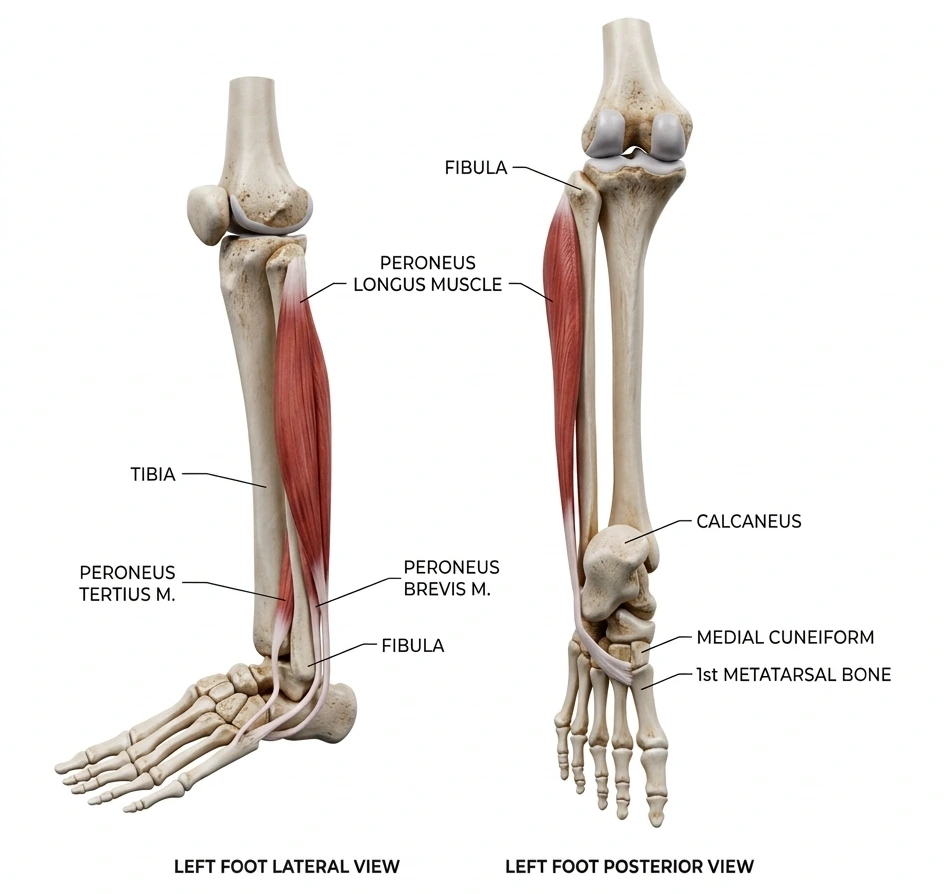
The peroneus longus muscle originates from the upper lateral aspect of the fibula and adjacent structures. Its muscle belly continues into a tendon that descends along the outer side of the lower leg.
The tendon passes behind the lateral malleolus, where it travels through the retromalleolar groove together with the peroneus brevis tendon. It then continues along the lateral aspect of the foot before passing beneath the cuboid and across the sole.
The tendon ultimately inserts on the base of the first metatarsal and medial cuneiform on the medial side of the foot.
Peroneus Brevis (Fibularis Brevis)
The peroneus brevis muscle originates from the lower part of the lateral surface of the fibula. Its tendon descends toward the ankle and passes behind the lateral malleolus.
At the ankle, the peroneus brevis tendon lies anterior to the peroneus longus tendon within the retromalleolar groove. Distal to the lateral malleolus, it continues toward the lateral aspect of the foot and inserts at the tuberosity at the base of the fifth metatarsal.
Peroneal Tendons at the Ankle
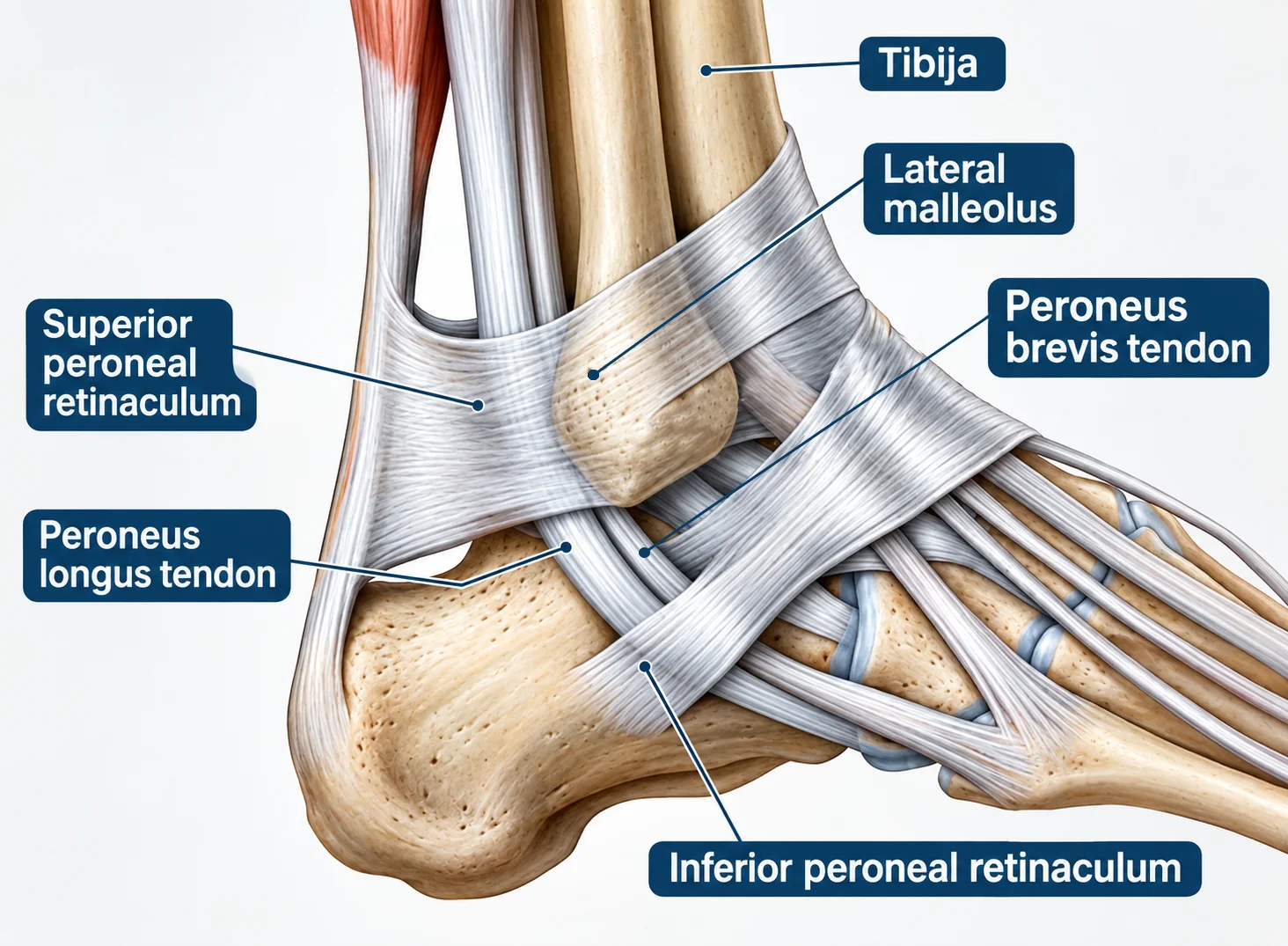
Around the lateral ankle, the peroneus longus and peroneus brevis tendons pass behind the lateral malleolus within a shallow retromalleolar groove. The peroneus brevis tendon lies anterior to the peroneus longus tendon at this level.
The tendons are held against the ankle by the superior and inferior peroneal retinacula. The superior peroneal retinaculum extends from the lateral malleolus toward the calcaneus and helps prevent the tendons from moving away from the fibula during ankle motion. More distally, the inferior peroneal retinaculum helps maintain the tendons in their anatomical position.
A synovial tendon sheath surrounds the tendons as they pass around the lateral ankle, reducing friction and allowing smooth movement. They share a common sheath in the retromalleolar region before following separate paths toward their insertions on the foot.
Disruption or laxity of the superior peroneal retinaculum can allow the tendons to move abnormally over the lateral malleolus, resulting in peroneal tendon subluxation or dislocation. Inflammation of the tendon sheath can cause peroneal tenosynovitis.
Function of the Peroneal Tendons
The main action of the peroneal tendons is eversion of the foot, which turns the sole outward. The peroneus longus also contributes to plantarflexion of the ankle and plays an important role in stabilizing the first ray and medial side of the foot.
Beyond producing movement, the peroneal tendons act as dynamic stabilizers of the lateral ankle. They help resist excessive inversion and respond rapidly to changes in foot position during walking, running, jumping, and other activities.
The peroneus longus and brevis therefore contribute to both movement and ankle stability, helping the foot adapt to changes in load and maintain controlled contact with the ground.
Common Peroneal Tendon Problems
Peroneal tendon disorders range from overuse-related tendon problems to acute tears, instability, and conditions affecting the os peroneum. Common problems include peroneal tendinopathy, tenosynovitis, tendon tears, subluxation or dislocation, and painful os peroneum syndrome.
1. Peroneal Tendinopathy
Peroneal tendinopathy refers to pain and structural changes affecting the peroneal tendons, usually as a result of repetitive loading or mechanical stress. It most commonly affects the peroneus longus and peroneus brevis tendons around the lateral ankle.
Symptoms typically include pain or tenderness along the outer side of the ankle, sometimes accompanied by swelling or discomfort during activities that load the peroneal tendons. Repetitive running, jumping, uneven surfaces, and previous ankle injuries may contribute to tendon overload.
The term peroneal tendinopathy is broader than peroneal tendonitis. Tendinopathy may involve a combination of tendon degeneration, altered tendon structure, and variable degrees of inflammation, whereas tendonitis specifically implies inflammation. In clinical practice, however, the terms are often used interchangeably.
For a more detailed discussion of symptoms, diagnosis, and treatment, see our guide to peroneal tendonitis.
2. Peroneal Tenosynovitis
Peroneal tenosynovitis is inflammation or irritation of the synovial sheath surrounding the peroneal tendons. Its causes are similar to those of peroneal tendinopathy, including repetitive loading, overuse, and ankle trauma.
Symptoms typically include pain and swelling around or behind the lateral malleolus, particularly with activities that load the peroneal tendons. Tendinopathy and tenosynovitis frequently occur together, and tendon tears can also be associated with inflammation of the surrounding tendon sheath.
3. Peroneal Tendon Tear
A peroneal tendon tear can range from a partial tear to a longitudinal split or complete rupture. The peroneus brevis is particularly prone to longitudinal split tears near the lateral malleolus, where the tendon is exposed to mechanical stress between the fibula and peroneus longus tendon.
Peroneal tendon tears may develop gradually from repetitive loading or occur after an ankle injury. Symptoms can include persistent pain behind the lateral malleolus, swelling, weakness with eversion, and sometimes a sensation of ankle instability.
Treatment depends on the extent of the tear, symptoms, tendon function, and activity level. Conservative treatment is generally the initial approach, while symptomatic tears that do not respond adequately to non-operative treatment may require surgery. When the tendon can be preserved, surgical debridement and repair or tubularization are preferred; irreparable tears may require tendon grafting or, in selected cases, tenodesis to the other peroneal tendon.
4. Peroneal Tendon Subluxation and Dislocation
Peroneal tendon subluxation or dislocation occurs when the peroneal tendons move abnormally out of the retromalleolar groove behind the lateral malleolus. It is commonly associated with an ankle injury, particularly a sudden forceful contraction of the peroneal muscles, and may also occur in athletes involved in running or rapid changes of direction.
Injury to the superior peroneal retinaculum (SPR) is an important factor because this structure normally keeps the tendons against the lateral malleolus. Patients may describe a characteristic snapping or popping sensation behind the lateral malleolus, sometimes accompanied by pain or a feeling of ankle instability.
Treatment depends on whether the instability is acute or chronic and on the patient’s activity level.
Acute instability in non-athletes may initially be treated with immobilization and rehabilitation, although recurrent instability can occur. Chronic instability and symptomatic cases that fail conservative treatment generally require surgical stabilization, while early surgical treatment is commonly recommended for athletes.
Surgical procedures typically focus on restoring the position of the tendons and repairing the superior peroneal retinaculum, with retromalleolar groove deepening often added in chronic cases and in athletes.
5. Os Peroneum and Peroneus Longus Tendon Disorders
The os peroneum is a small accessory ossicle located within the peroneus longus tendon, usually near the cuboid bone on the lateral aspect of the foot. It is a relatively common anatomical finding and is often asymptomatic and discovered incidentally on imaging.
However, the os peroneum can also be associated with disorders of the peroneus longus tendon. Trauma or repetitive loading may cause pain around the lateral midfoot and can affect both the tendon and the ossicle.
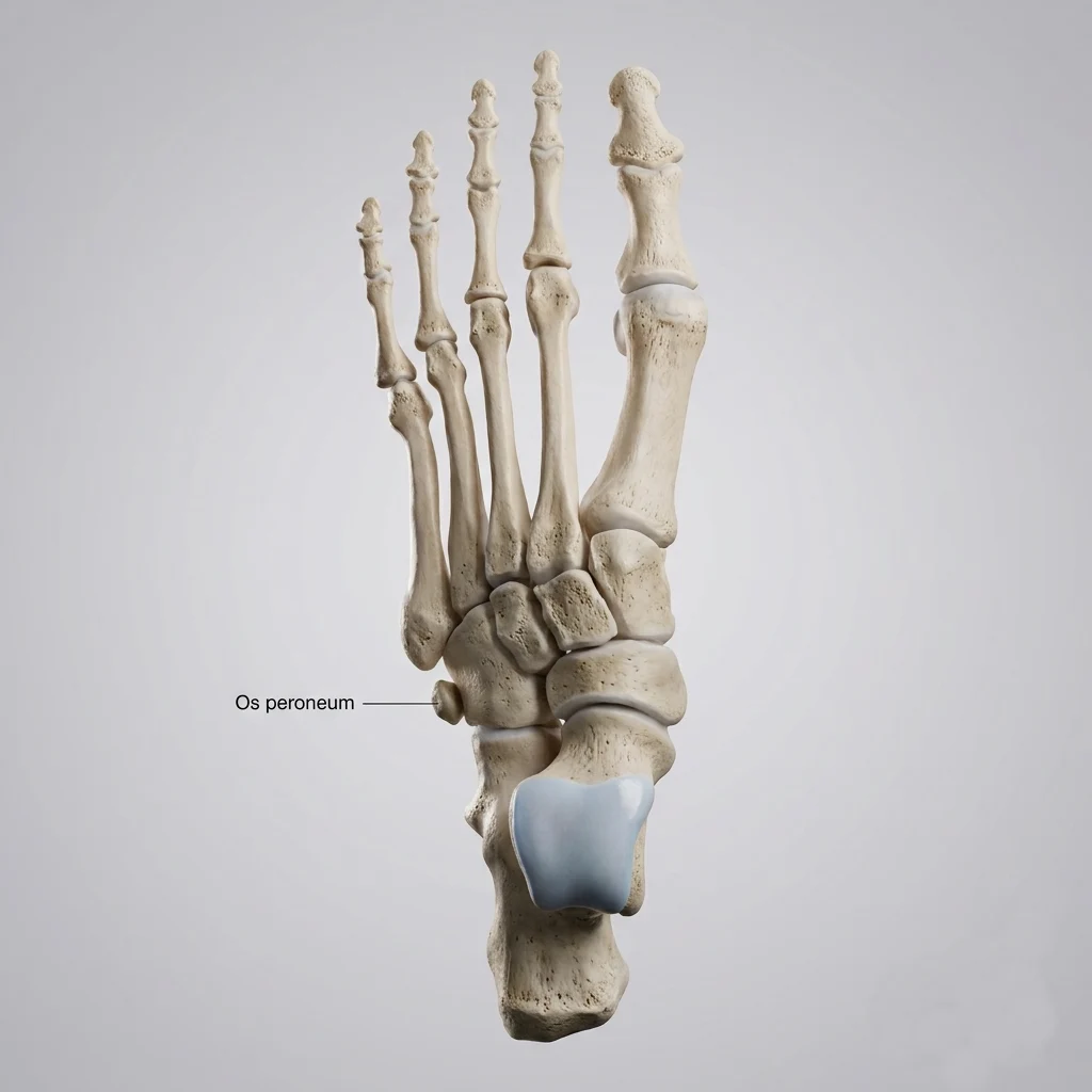
A fractured or displaced os peroneum may be particularly important clinically. These findings can indicate a significant injury to the peroneus longus tendon, including a partial or complete tendon tear. Therefore, an abnormal os peroneum should be assessed together with the surrounding tendon rather than considered an isolated bone abnormality.
When symptoms are present, the combination of lateral foot pain and abnormalities of the os peroneum may be referred to as painful os peroneum syndrome (POPS). Ultrasound and MRI can help evaluate the os peroneum and determine whether associated peroneus longus tendon pathology is present.
Symptoms of Peroneal Tendon Disorders
Peroneal tendon disorders usually cause pain along the outer side of the ankle or foot. Symptoms can vary depending on which tendon is affected and whether the problem involves tendinopathy, tenosynovitis, a tendon tear, or tendon instability.
Common symptoms include:
- Pain behind or below the lateral malleolus
- Swelling around the outer ankle
- Tenderness along the peroneal tendons
- Pain with eversion of the foot
- Weakness during eversion or other activities that load the tendons
- Snapping or popping sensation behind the lateral malleolus
- Feeling of ankle instability, particularly during walking or sports
Location of Pain
The location of pain can provide useful clues about the structures involved, although symptoms often overlap between different peroneal tendon disorders.
| Pain Location | Possible Cause |
|---|---|
| Behind the lateral malleolus | Peroneal tendinopathy, tenosynovitis, or tendon tear |
| Below the lateral malleolus | Peroneal tendon pathology or irritation of the tendons as they curve around the ankle |
| Along the lateral side of the foot | Peroneus brevis tendon pathology or pain related to its insertion at the base of the fifth metatarsal |
| Lateral midfoot near the cuboid | Peroneus longus tendon pathology or painful os peroneum syndrome |
| Snapping behind the lateral malleolus | Peroneal tendon subluxation or dislocation |
How Are Peroneal Tendon Problems Diagnosed?
Diagnosis of a peroneal tendon disorder usually starts with a clinical examination. Imaging can then help confirm the diagnosis, determine the extent of tendon damage, and identify associated abnormalities.
Clinical Examination
The examination focuses on the location of pain, tendon function, and ankle stability. The clinician may assess:
- Palpation along the peroneal tendons, particularly behind and below the lateral malleolus
- Resisted eversion to reproduce pain or assess tendon strength
- Single-leg stance to assess ankle control and stability
- Circumduction of the ankle to identify pain, weakness, or tendon snapping
- Assessment of ankle instability, particularly after previous ankle sprains
A snapping sensation during ankle movement may raise suspicion of peroneal tendon subluxation or dislocation.
Ultrasound
Musculoskeletal ultrasound is particularly useful for evaluating the peroneal tendons because it allows the tendons to be examined in real time and compared with the opposite side.
Ultrasound can identify:
- Tendon thickening and structural abnormalities associated with tendinopathy
- Tenosynovitis and fluid within the tendon sheath
- Partial and longitudinal split tears
- Tendon subluxation or dislocation
- Changes in the peroneus longus and peroneus brevis tendons during movement
One of the major advantages of ultrasound is dynamic assessment. The peroneal tendons can be observed while the ankle is moved, particularly as they pass behind the lateral malleolus. This can reveal intermittent subluxation or dislocation that may not be apparent during a static examination.
MRI
MRI provides detailed visualization of the peroneal tendons and surrounding soft tissues. It can help evaluate:
- Partial or complete tendon tears
- Tendon degeneration and surrounding inflammation
- The tendon sheath and retinacula
- Associated bone and osteochondral abnormalities
- Other soft-tissue conditions that may contribute to lateral ankle pain
Ultrasound and MRI are complementary rather than interchangeable. Dynamic ultrasound is particularly valuable when tendon instability is suspected, while MRI can provide a broader assessment of the tendon and surrounding structures.
Treatment of Peroneal Tendon Disorders
Treatment depends on the specific tendon problem, its severity, symptoms, and the patient’s activity level. Many peroneal tendon disorders can initially be managed without surgery.
Conservative Treatment
Conservative treatment may include:
- Activity modification to reduce painful loading
- Relative rest and short-term immobilization when symptoms are severe
- Physical therapy to restore tendon strength, ankle mobility, and stability
- Progressive strengthening, particularly of the peroneal muscles
- Treatment of contributing factors, such as recurrent ankle instability or abnormal foot mechanics
Surgical Treatment
Surgery may be considered for complete or significant tendon tears, persistent symptoms despite appropriate conservative treatment, or chronic tendon subluxation or dislocation. Depending on the underlying problem, procedures may include tendon repair or tubularization, tenodesis, tendon grafting, or stabilization of the peroneal tendons and retinaculum.
When Should You See a Doctor?
You should consider medical evaluation if you develop:
- Persistent pain along the outer ankle or foot
- Pain after an acute ankle injury, particularly if symptoms do not improve
- Snapping or popping behind the lateral malleolus
- Persistent swelling around the outer ankle
- Weakness with foot eversion
- Difficulty or inability to bear weight
- A feeling of ankle instability during walking or sports
Early assessment can help identify a tendon tear or instability and guide appropriate treatment.
Bottom Line
The peroneal tendons are important dynamic stabilizers of the lateral ankle. Problems range from overuse-related tendinopathy and tenosynovitis to tendon tears, painful os peroneum, and tendon instability. A clinical examination combined with ultrasound or MRI when appropriate can help determine the underlying problem and guide treatment.
van Dijk PA, Miller D, Calder J, DiGiovanni CW, Kennedy JG, Kerkhoffs GM, Kynsburtg A, Havercamp D, Guillo S, Oliva XM, Pearce CJ, Pereira H, Spennacchio P, Stephen JM, van Dijk CN. The ESSKA-AFAS international consensus statement on peroneal tendon pathologies. Knee Surg Sports Traumatol Arthrosc. 2018 Oct;26(10):3096-3107. PubMed: International consensus on peroneal tendon pathologies
Dombek MF, Orsini RC, Mendicino RW, Saltrick KR. Peroneal tendon tears: a retrospective review. J Foot Ankle Surg. 2003;42(5):250-258. PubMed: Peroneal tendon tears and clinical management
van Dijk PA, Kerkhoffs GM, van Dijk CN. Peroneal tendon disorders: an overview. Foot Ankle Clin. PubMed: Overview of peroneal tendon disorders
Bianchi S, Martinoli C. Ultrasound of the musculoskeletal system: peroneal tendons and ankle. Springer. Clinical reference: Musculoskeletal ultrasound of the ankle and peroneal tendons
Brigido MK, Fessell DP, Jacobson JA, Widman G, Craig JG, Jamadar DA, Hayes CW. Radiologic evaluation of the ankle tendons: ultrasound and MRI. Radiographics. PubMed: Imaging evaluation of ankle tendons
Brigido MK, Fessell DP, Jacobson JA, Widman G, Craig JG, Jamadar DA, Hayes CW. Ultrasound and MRI of the peroneal tendons. Clinical imaging reference: Ultrasound and MRI assessment of peroneal tendon pathology