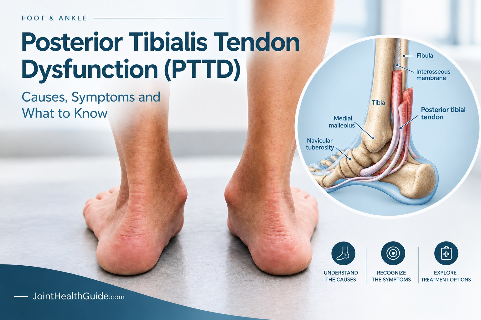
Posterior tibial tendon dysfunction (PTTD) is a common cause of inner ankle pain and progressive flattening of the foot arch. The condition develops when the posterior tibial tendon becomes irritated, weakened, or damaged and can no longer provide adequate support to the foot.
Many people first notice pain on the inside of the ankle, swelling, or discomfort during walking and exercise. In the early stages, symptoms may resemble posterior tibial tendonitis or tibialis posterior tendonitis, but untreated tendon damage can eventually lead to adult-acquired flatfoot deformity.
Early diagnosis is important because treatment is often most effective before significant structural changes occur. Healthcare providers commonly classify the condition into four posterior tibial tendon dysfunction stages, ranging from mild tendon irritation to advanced foot and ankle deformity.
This article explains the symptoms, causes, stages, diagnosis, treatment options, and recovery process for posterior tibial tendon dysfunction.
Key Clinical Points – Posterior Tibial Tendon Dysfunction (PTTD)
- Posterior tibial tendon dysfunction (PTTD) is a common cause of medial ankle pain and acquired flatfoot in adults.
- Typical symptoms include pain and swelling along the inner ankle, weakness during push-off, and progressive flattening of the medial longitudinal arch.
- The “too many toes” sign indicates forefoot abduction and may be present in more advanced flexible deformity.
- Diagnosis is based on clinical examination and weight-bearing radiographs, with ultrasound or MRI used when further assessment of the tendon is needed.
- Treatment depends on disease stage and may include activity modification, supportive footwear, orthoses, physical therapy, and progressive strengthening exercises.
- Early diagnosis and treatment can relieve symptoms and help prevent progression to rigid flatfoot deformity and arthritis. Advanced cases may require surgery.
What Is Posterior Tibial Tendon Dysfunction?
Posterior tibial tendon dysfunction (PTTD) is a progressive condition that affects the posterior tibial tendon, one of the primary structures responsible for supporting the arch of the foot. The disorder develops when the tendon becomes irritated, weakened, or damaged and can no longer perform its normal function effectively. Over time, this loss of tendon function may lead to pain, flattening of the foot arch, altered walking mechanics, and, in more advanced cases, adult-acquired flatfoot deformity.
PTTD often begins gradually. Many people initially experience mild discomfort along the inside of the ankle, especially after prolonged walking, running, or standing. As the condition progresses, the tendon may lose its ability to stabilize the foot, causing the arch to collapse and the foot to roll inward excessively.
What Does the Posterior Tibial Tendon Do?
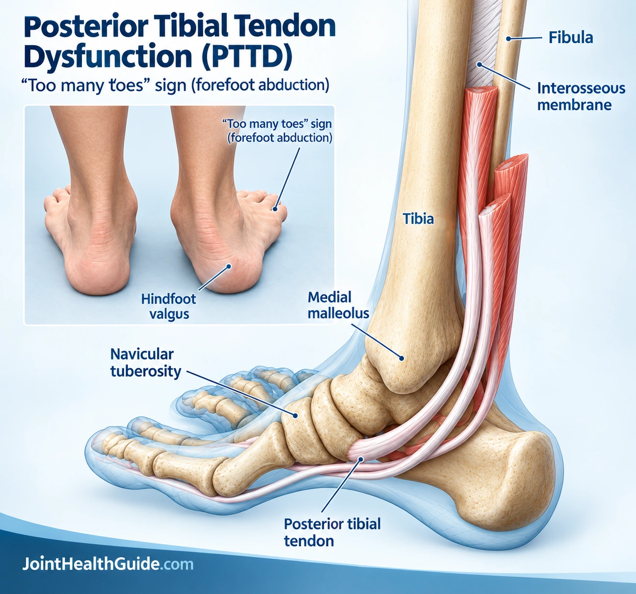
The posterior tibial tendon connects the tibialis posterior muscle in the calf to several bones on the inside of the foot. It plays a critical role in maintaining normal foot mechanics during walking and other weight-bearing activities.
Its primary functions include:
- Supporting the medial longitudinal arch of the foot.
- Controlling foot pronation during walking and running.
- Helping stabilize the foot during the push-off phase of gait.
- Assisting with inversion, the motion that turns the foot inward.
When the tendon is healthy, it helps distribute forces efficiently throughout the foot and ankle. However, when it becomes weakened or damaged, the arch can gradually flatten, placing additional stress on surrounding joints, ligaments, and muscles.
Is PTTD the Same as Posterior Tibial Tendonitis?
Not exactly. Although the terms are often used interchangeably, they describe different stages or aspects of the same problem.
Posterior tibial tendonitis and tibialis posterior tendonitis typically refer to an inflammatory reaction within the tendon. This phase is often associated with recent overuse, increased physical activity, or a sudden rise in training volume. Symptoms commonly include pain, tenderness, and mild swelling along the inside of the ankle.
In contrast, tibialis posterior tendinopathy refers to chronic tendon degeneration. Rather than active inflammation, the tendon develops structural changes that reduce its strength and ability to tolerate load. This is the most common underlying process in long-standing cases.
Posterior tibial tendon dysfunction (PTTD) is the broader clinical condition that results when tendon damage begins to affect foot function. As the tendon loses strength, it may no longer adequately support the arch, leading to progressive deformity, altered gait mechanics, and worsening symptoms.
In simple terms:
- Tendonitis = predominantly inflammatory stage.
- Tendinopathy = degenerative tendon changes.
- PTTD = functional impairment caused by tendon damage and weakening.
Understanding this distinction is important because treatment strategies often depend on whether the tendon is primarily inflamed, degenerative, or already causing structural changes in the foot.
Symptoms of Posterior Tibial Tendon Dysfunction
The symptoms of posterior tibial tendon dysfunction (PTTD) usually develop gradually and tend to worsen over time. Many people first notice pain on the inside of the ankle, especially during long walks, exercise, or prolonged standing. The discomfort may start as a mild ache but can become more persistent as the tendon becomes weaker.
A common early symptom is inner ankle pain located just behind and below the bony prominence on the inside of the ankle. The area may feel tender to the touch, and some people notice mild swelling along the course of the tendon. At this stage, symptoms are often mistaken for a simple strain or posterior tibial tendonitis.

As the condition progresses, routine activities such as climbing stairs, walking on uneven surfaces, or standing on tiptoes may become more difficult. Some individuals report fatigue in the foot or ankle and feel that the affected foot is becoming weaker or less stable during walking.
Over time, the arch of the foot may begin to flatten. As this happens, the foot often rolls inward while the forefoot drifts outward. Shoes may fit differently, and you may notice increased wear on the inner side of the sole. Pain can also spread from the inner ankle to the arch of the foot and, in more advanced cases, to the outer side of the ankle as joint alignment changes.
Common symptoms of PTTD include:
- Painful inner ankle that worsens with activity
- Swelling along the inside of the ankle
- Tenderness over the posterior tibial tendon
- Pain in the arch of the foot
- Difficulty standing on tiptoes on the affected side
- Foot fatigue during walking or exercise
- Progressive flattening of the foot arch
- A feeling of instability or weakness in the foot
PTTD typically progresses through several stages, with symptoms becoming more noticeable as tendon function declines.
Recognizing the condition early and seeking treatment before significant arch collapse occurs can help improve outcomes and reduce the risk of long-term foot deformity.
What Causes Posterior Tibial Tendon Dysfunction?
Posterior tibial tendon dysfunction (PTTD) usually develops as a result of gradual wear and tear rather than a single injury. Repetitive stress placed on the tendon over many years can lead to small areas of damage that accumulate over time. As the tendon weakens, it becomes less able to support the arch of the foot, increasing the risk of pain, flatfoot deformity, and progressive loss of function.
Several factors can increase the likelihood of developing PTTD. Excess body weight is one of the most important risk factors because it places additional strain on the foot and ankle with every step. Research has shown that overweight and obesity are extremely common among people diagnosed with the condition. Posterior tibial tendon dysfunction is more common in women, particularly after the age of 50.
Certain medical conditions may further increase risk. These include diabetes, high blood pressure, elevated cholesterol levels, and inflammatory diseases that can affect tendon health and healing capacity. Previous ankle injuries, longstanding flat feet, and occupations or activities that involve prolonged standing or walking may also contribute to tendon degeneration over time.
Although PTTD is often considered a tendon disorder, experts now recognize that the condition can involve other structures that help support the arch, including ligaments and joints in the foot. This is one reason why symptoms may gradually worsen and why some people develop progressive flatfoot deformity even when tendon damage alone does not fully explain their condition.
How Is Posterior Tibial Tendon Dysfunction Diagnosed?
The diagnosis of posterior tibial tendon dysfunction (PTTD) usually begins with a review of your symptoms and a physical examination. In many cases, doctors can suspect the condition based on the location of your pain, changes in foot shape, and how your foot functions during standing and walking.
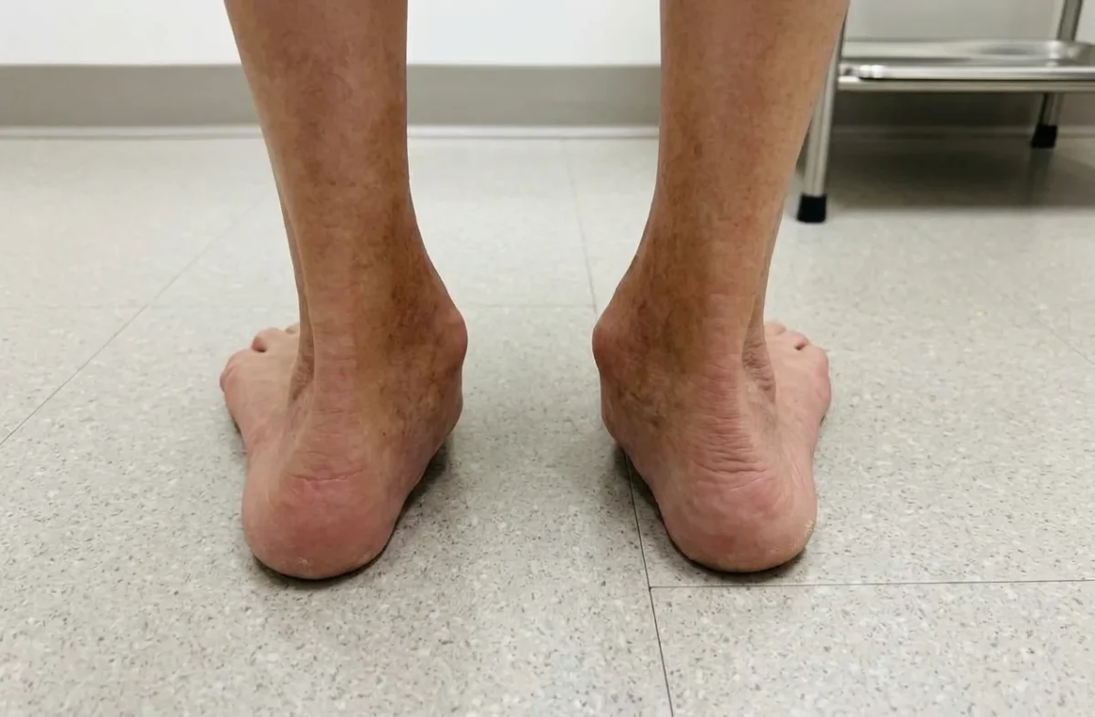
During the examination, your healthcare provider will look for tenderness and swelling along the posterior tibial tendon, which runs behind the inner ankle bone and into the arch of the foot. They will also assess the position of your foot while standing. One of the most recognizable findings is the too many toes sign. When viewed from behind, more toes than normal are visible on the affected side because the forefoot drifts outward as the arch collapses.
Your doctor may also ask you to perform a single-leg heel raise, which involves standing on one foot and rising onto your toes. Difficulty performing this movement, pain during the test, or an inability to lift the heel can indicate weakness or dysfunction of the posterior tibial tendon.
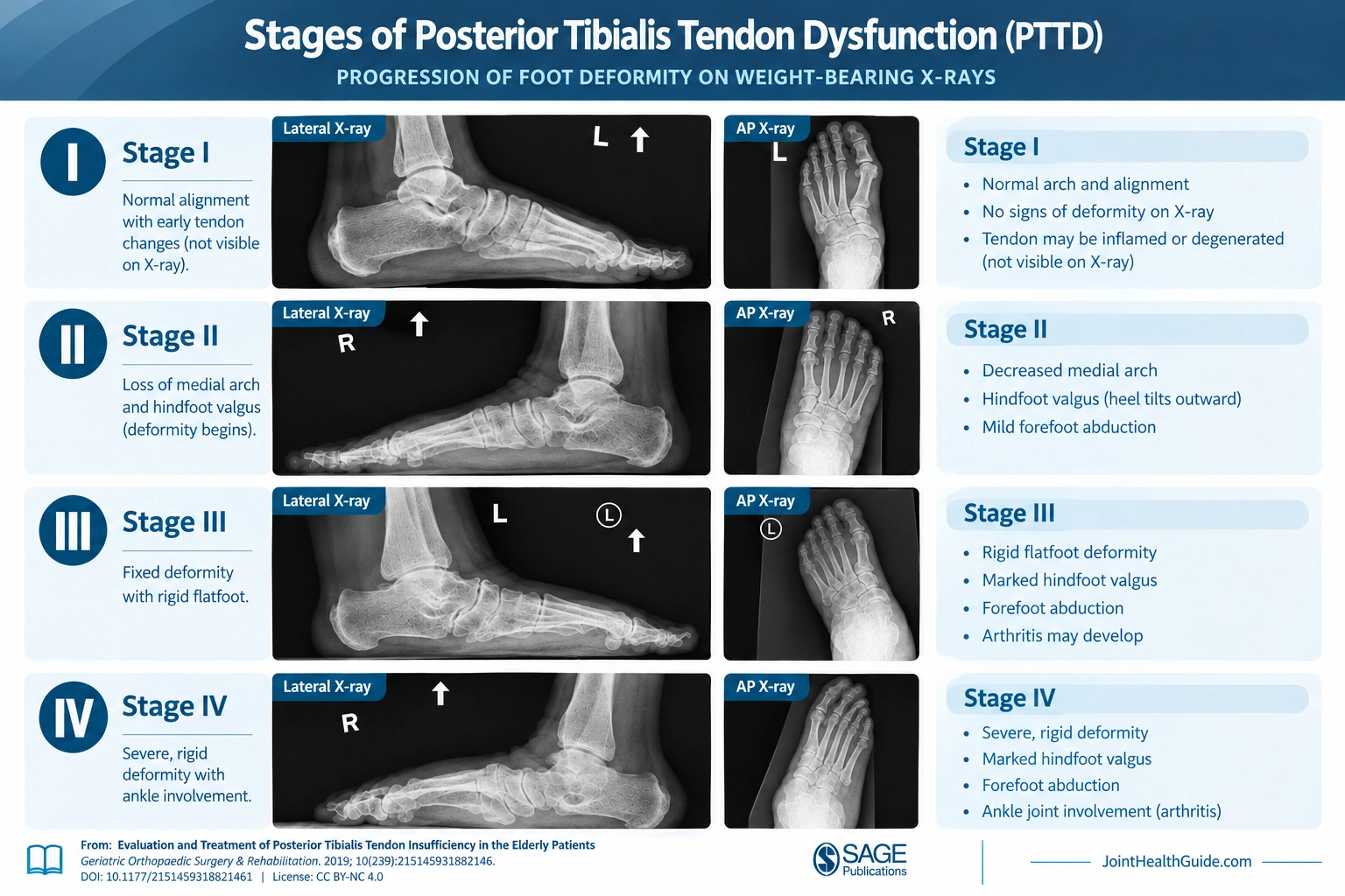
Imaging studies are often used to confirm the diagnosis and evaluate the severity of the condition. Weight-bearing X-rays can reveal flattening of the arch, changes in foot alignment, and other signs of progressive flatfoot deformity. While X-rays do not show the tendon itself, they help assess the structural effects of long-standing PTTD.
Ultrasound can directly visualize the tendon and may show tendon thickening, inflammation consistent with posterior tibial tendonitis, or signs of chronic degeneration. It is often used as a quick and cost-effective imaging option.
MRI provides the most detailed evaluation of the tendon and surrounding soft tissues. It can help identify tendon tears, advanced tibialis posterior tendinopathy, ligament injuries, and other abnormalities that may contribute to symptoms.
Clinical Insight
Combining the patient’s history, physical examination findings, and appropriate imaging studies allows healthcare providers to diagnose PTTD accurately and determine its stage, which helps guide treatment decisions.
Posterior Tibial Tendon Dysfunction Stages
Healthcare providers commonly classify posterior tibial tendon dysfunction into four stages based on the severity of tendon damage, the degree of arch collapse, and the flexibility of the foot deformity. Staging is important because treatment recommendations and expected outcomes often vary depending on how far the condition has progressed.
In the early stages, the tendon remains functional and the foot retains its normal shape. As the condition advances, the tendon becomes less capable of supporting the arch, leading to progressive flattening of the foot and changes in alignment. In the most advanced stages, the deformity becomes rigid and may begin to affect the ankle joint itself.
The table below summarizes the key features of each stage of PTTD:
| Stage | Arch collapse | Flexibility | Key findings |
|---|---|---|---|
| Stage I | Absent | Normal foot alignment | Mild tenosynovitis; single-leg heel raise preserved; normal radiographs. |
| Stage IIA | Present | Flexible | Flatfoot deformity; inability to perform a single-leg heel raise. |
| Stage IIB | Present | Flexible | Forefoot abduction, positive “too many toes” sign, and greater than 40% talonavicular uncoverage. |
| Stage III | Present | Rigid | Fixed flatfoot deformity, hindfoot valgus, and subtalar arthritis. |
| Stage IV | Present | Rigid | Rigid flatfoot, hindfoot valgus, deltoid ligament insufficiency, and valgus talar tilt at the ankle; subtalar arthritis may coexist. |
Conditions That Can Cause Similar Inner Ankle Pain
Posterior tibial tendon dysfunction is a common cause of pain along the inside of the ankle, but several other conditions can produce similar symptoms. Because treatment varies depending on the underlying cause, obtaining an accurate diagnosis is important.
Other conditions that may cause inner ankle pain include:
- Deltoid ligament injury – Damage to the deltoid ligament on the inside of the ankle can cause pain, swelling, and tenderness that may resemble PTTD. Symptoms often develop after an ankle twist or traumatic injury.
- Tarsal tunnel syndrome – This condition occurs when the tibial nerve becomes compressed as it passes through the tarsal tunnel near the inner ankle. In addition to pain, people may experience burning, tingling, numbness, or electric shock-like sensations that radiate into the foot.
- Flexor tendon injuries – Tendons such as the flexor hallucis longus and flexor digitorum longus also travel behind the inner ankle. Tendinopathy or inflammation affecting these tendons can produce symptoms similar to posterior tibial tendon dysfunction.
- Medial ankle sprain – Although less common than lateral ankle sprains, injuries to the structures on the inside of the ankle can cause persistent pain, swelling, and instability.
- Ankle osteoarthritis – Degenerative changes within the ankle joint may result in chronic pain, stiffness, swelling, and reduced mobility. Symptoms are often worse after prolonged activity or during the first few steps after resting.
- Spring ligament injury – The spring ligament helps support the arch of the foot and often works together with the posterior tibial tendon. Injury to this ligament may contribute to arch collapse and symptoms that closely resemble PTTD.
Because several disorders can present with a painful inner ankle, a thorough physical examination and, when appropriate, imaging studies are often necessary to determine the exact cause of symptoms and guide treatment.
Treatment Options for PTTD
Treatment for posterior tibial tendon dysfunction depends largely on the stage of the condition.
Early-stage PTTD often responds well to conservative treatment, while advanced cases with significant deformity may require surgery.
The primary goals of treatment are to reduce pain, support the arch, improve foot function, and prevent further progression of the condition.
Conservative Treatment
Conservative treatment is typically the first-line approach for Stage I and Stage II PTTD. These stages are characterized by tendon dysfunction without severe rigid deformity, making nonsurgical interventions particularly effective.
Treatment usually begins with activity modification to reduce stress on the posterior tibial tendon. Patients are often advised to temporarily limit activities that aggravate symptoms, such as running, prolonged walking, or high-impact exercise. Nonsteroidal anti-inflammatory drugs (NSAIDs) may help reduce pain, especially during periods of increased tendon irritation.
In patients with significant pain, short-term immobilization in a walking boot may be recommended for several weeks to allow the tendon to recover. Once symptoms begin to improve, physical therapy becomes a key component of treatment. Rehabilitation programs typically focus on improving ankle mobility, calf flexibility, balance, and progressive strengthening of the tibialis posterior muscle and other structures that support the arch.
Orthotics and ankle braces are frequently prescribed to reduce strain on the tendon and improve foot alignment. Custom arch supports may be particularly helpful in patients with flexible flatfoot deformity. Long-term use of supportive footwear and orthotics can help reduce symptoms and slow disease progression.
Weight management should also be considered when appropriate, as excess body weight places additional stress on the tendon with every step.
Many patients diagnosed in Stage I or Stage II experience significant improvement with a comprehensive conservative treatment program followed consistently for several months.
Surgical Treatment
Surgery may be considered when symptoms persist despite appropriate conservative treatment or when the condition has progressed to more advanced stages.
In Stage I and early Stage II PTTD, surgical procedures generally focus on repairing or reconstructing the damaged tendon while correcting mild alignment abnormalities. Tendon debridement, tendon repair, or tendon transfer procedures may be combined with corrective osteotomies to restore foot mechanics.
In Stage III PTTD, the deformity becomes more rigid and arthritis may develop in the hindfoot joints. As a result, joint fusion procedures are often required to stabilize the foot and relieve pain.
In Stage IV PTTD, the deformity extends to the ankle joint. Treatment may require more extensive reconstructive procedures, ligament reconstruction, ankle realignment, or selected fusion procedures depending on the severity of the deformity and joint involvement.
While surgery can provide good outcomes in appropriately selected patients, most individuals diagnosed in the early stages can often avoid surgery through timely diagnosis, structured rehabilitation, appropriate bracing, and long-term management of risk factors.
Best Exercises for Posterior Tibial Tendon Dysfunction
Calf Stretch
- Purpose: Improves calf flexibility and reduces restricted ankle dorsiflexion.
- How to perform: Stand facing a wall with the affected leg behind you. Keep the heel on the floor and the knee straight while leaning forward.
- Repetitions: Hold for 30 seconds; repeat 3 times.
Heel Raises
- Purpose: Strengthens the calf muscles and posterior tibial tendon.
- How to perform: Stand with feet hip-width apart and slowly rise onto your toes. Lower your heels in a controlled manner. Begin with double-leg raises and progress to single-leg raises as tolerated.
- Repetitions: 2–3 sets of 8–12 repetitions.
Resisted Foot Inversion
- Purpose: Strengthens the posterior tibial muscle and improves medial ankle support.
- How to perform: Sit with your leg extended and a resistance band around the forefoot. Turn the foot inward against the band’s resistance while keeping the leg still.
- Repetitions: 2–3 sets of 10–15 repetitions.
Balance Exercises
- Purpose: Improves balance, proprioception, and lower-limb stability.
- How to perform: Stand on the affected leg while maintaining a stable foot position. Use a wall or chair for support if needed.
- Repetitions: Hold for 20–30 seconds; repeat 3 times.
Foot Intrinsic Muscle Strengthening
- Purpose: Strengthens the intrinsic foot muscles and supports the medial longitudinal arch.
- How to perform: Perform the short-foot exercise by gently drawing the ball of the foot toward the heel without curling the toes or lifting the heel.
- Repetitions: Hold for 5–10 seconds; repeat 10 times.
Recovery Time and Prognosis
How Long Does PTTD Take to Heal?
Early-stage PTTD may improve within 6–12 weeks with appropriate treatment, although recovery can take several months. Advanced cases often require longer treatment.
Can Posterior Tibial Tendon Dysfunction Be Reversed?
Early symptoms and inflammation can improve with conservative treatment. However, established tendon degeneration and structural foot deformity may not be fully reversible.
What Happens Without Treatment?
PTTD may progress to progressive arch collapse, rigid flatfoot deformity, chronic pain, and arthritis, potentially requiring surgery.
How to Prevent Posterior Tibial Tendon Dysfunction
Although PTTD cannot always be prevented, several measures may reduce tendon overload and help maintain foot function.
- Proper footwear: Wear supportive shoes that provide adequate arch support and stability.
- Gradual training progression: Increase exercise intensity and walking or running distance gradually.
- Weight management: Maintain a healthy body weight to reduce stress on the posterior tibial tendon.
- Early treatment of tendon pain: Address persistent medial ankle pain or swelling before symptoms progress.
- Strength training: Regularly strengthen the calf muscles and intrinsic foot muscles to support foot function.
Frequently Asked Questions
Is posterior tibial tendon dysfunction serious?
PTTD can become serious if left untreated. Progressive tendon dysfunction may lead to arch collapse, rigid flatfoot deformity, and arthritis.
Can PTTD heal without surgery?
Yes. Early-stage PTTD often improves with conservative treatment, including activity modification, orthoses, physical therapy, and targeted exercises. Advanced deformity may require surgery.
What are the stages of posterior tibial tendon dysfunction?
The Johnson and Strom classification, modified by Myerson, describes stages I–IV, ranging from tendon inflammation without deformity to rigid flatfoot with ankle involvement.
What is the too many toes sign?
The “too many toes” sign occurs when several toes are visible from behind the foot due to forefoot abduction. It is commonly associated with stage IIB PTTD.
Can walking make PTTD worse?
Yes. Prolonged walking or excessive loading can aggravate pain and swelling, particularly without adequate support. Activity should be adjusted to symptom severity.
Is PTTD the same as posterior tibial tendonitis?
No. Tendonitis refers to tendon inflammation, whereas PTTD encompasses a broader spectrum of tendon dysfunction, degeneration, and associated foot deformity.
What is the best exercise for PTTD?
Progressive heel raises are commonly used to strengthen the posterior tibial tendon and calf muscles. The appropriate exercise and intensity depend on disease stage and symptom severity.
Can PTTD cause flat feet?
Yes. Dysfunction of the posterior tibial tendon can compromise medial arch support, leading to progressive arch collapse and acquired flatfoot deformity.
Conclusion
Posterior tibial tendon dysfunction (PTTD) is a common cause of medial ankle pain and acquired flatfoot in adults. Early recognition of symptoms and timely treatment are essential to preserve tendon function and foot alignment. Appropriate conservative management can relieve symptoms and help slow or prevent progression of the deformity, potentially avoiding more complex surgical treatment.
Knapp PW, Constant D. Posterior Tibial Tendon Dysfunction (Archived). StatPearls [Internet]. Updated May 21, 2024. NCBI Bookshelf: Clinical overview, staging, diagnosis, and treatment of PTTD
Ikpeze TC, Brodell JD Jr, Chen RE, Oh I. Evaluation and Treatment of Posterior Tibialis Tendon Insufficiency in the Elderly Patients. Geriatr Orthop Surg Rehabil. 2019;10:2151459318821461. doi:10.1177/2151459318821461. Full-text review: Evaluation and treatment of posterior tibial tendon insufficiency
Ling SK, Lui TH. Posterior Tibial Tendon Dysfunction: An Overview. Open Orthop J. 2017;11:714–723. doi:10.2174/1874325001711010714. Full-text review: PTTD pathophysiology, clinical presentation, classification, and management