
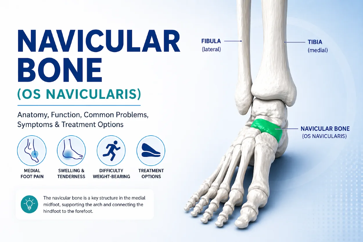
The navicular bone (also known as the os navicularis) is a small but essential bone located on the inner side of the midfoot. It plays a central role in maintaining the arch of the foot and ensuring smooth, stable movement during walking, running, and standing.
Because of its position between the ankle and the forefoot, the navicular bone acts as a key stabilizer and force distributor. When this bone is affected by injury or structural issues, it can lead to pain along the inner foot and impact overall mobility.
Despite its importance, the navicular bone is often overlooked until problems arise—making it crucial to understand its anatomy, function, and the most common conditions associated with it.
Key Clinical Points – Navicular Bone (Os Navicularis)
- The navicular bone (os navicularis) is a central structure in the medial midfoot, essential for maintaining the foot arch and stability.
- It plays a key role in load distribution and connects the ankle (talus) to the forefoot via the cuneiform bones.
- The navicular is a critical attachment point for the posterior tibial tendon, which supports the medial longitudinal arch.
- Common conditions include navicular stress fractures, accessory navicular syndrome, and posterior tibial tendon dysfunction.
- Typical symptoms involve medial foot pain, swelling, and difficulty with weight-bearing, especially during activity.
- Most cases respond well to conservative treatment, including rest, orthotics, and physical therapy, when diagnosed early.
What Is the Navicular Bone?
The navicular bone (also known as the os navicularis) is a small, boat-shaped bone located on the inner (medial) side of the midfoot. It is one of the tarsal bones and plays a key role in connecting the ankle to the front of the foot. The term os navicularis comes from Latin, meaning “little boat,” which reflects its curved shape.
Positioned between the talus and the cuneiform bones, the navicular bone acts as an important structural link within the foot. It contributes to stability, helps distribute forces during movement, and supports the medial arch.
Despite its small size, the navicular bone is essential for normal foot function, particularly during walking and running.
Anatomy of the Navicular Bone
The navicular bone (os navicularis) has a unique structure that reflects its important role in foot stability and movement. Its shape, location, and connections with surrounding bones make it a central component of the medial midfoot.
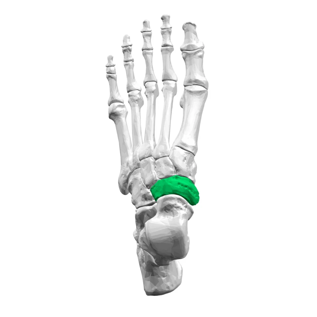
Shape and Structure
The navicular bone has a curved, boat-like shape, which is where its name originates (from the Latin navicula, meaning “small boat”). It features multiple articular surfaces covered with cartilage, allowing smooth movement between adjacent bones.
Although relatively small, the navicular bone is structurally strong and designed to withstand significant forces during weight-bearing activities such as walking and running.
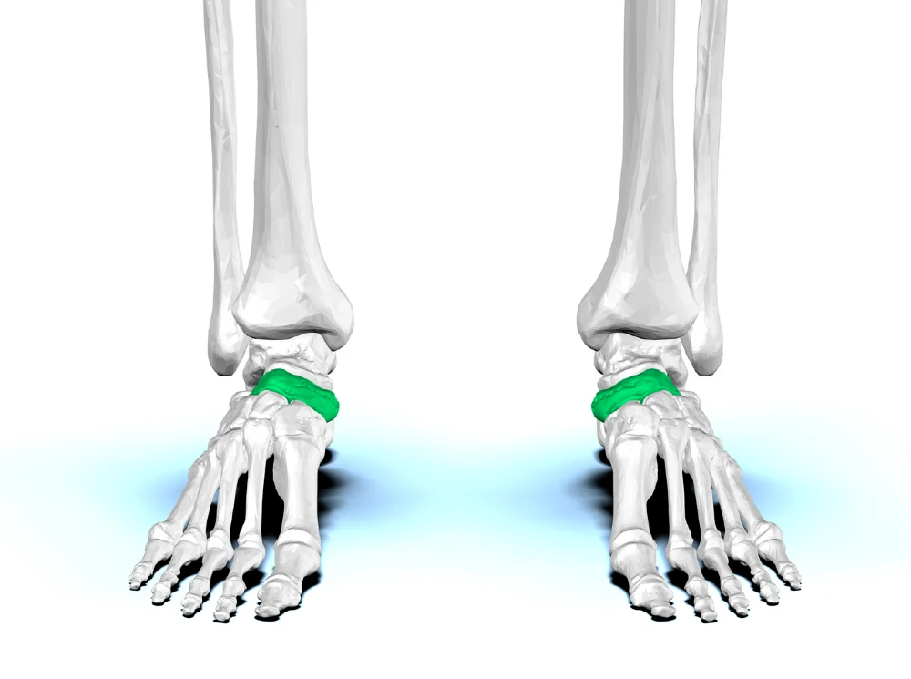
Location in the Foot
The navicular bone is located on the medial (inner) side of the midfoot. It sits between the rearfoot and forefoot, forming a key transition point for force transmission.
Because of this position, the navicular bone plays a central role in maintaining the alignment of the foot and supporting the medial longitudinal arch.
Articulations (Talus and Cuneiform Bones)
The navicular bone forms joints with several surrounding bones:
- Talus – the navicular articulates proximally with the talus, forming the talonavicular joint, which is essential for foot mobility
- Three cuneiform bones – distally, it connects with the medial, intermediate, and lateral cuneiforms, linking it to the forefoot
These articulations allow the foot to remain both flexible and stable during movement, adapting to different surfaces and loads.
Navicular Tuberosity and Tendon Attachment
One of the most important features of the navicular bone is the navicular tuberosity, a bony prominence located on its medial side.
This structure serves as the primary attachment point for the posterior tibial tendon, which plays a crucial role in:
- Supporting the arch of the foot
- Stabilizing the midfoot
- Controlling foot movement during walking
Clinical Insight
Dysfunction or stress in this area—particularly involving the posterior tibial tendon—can lead to pain and conditions such as accessory navicular syndrome or progressive arch collapse.
Function of the Navicular Bone
The navicular bone (os navicularis) plays a central role in foot mechanics by acting as a key stabilizing structure in the midfoot. It is essential for maintaining proper alignment of the medial arch and ensuring efficient force transfer between the rearfoot and forefoot during movement.
Key Functions:
- Maintains the medial longitudinal arch
The navicular bone is a critical support point for the medial arch, helping preserve the natural curvature of the foot. - Facilitates load distribution and biomechanics
It helps transfer and distribute body weight and mechanical forces from the talus to the forefoot during standing and movement. - Supports efficient gait mechanics
By acting as a central link in the midfoot, it contributes to smooth transitions during walking, running, and jumping. - Provides tendon leverage and stability
Serves as an attachment site for the posterior tibial tendon, which actively supports arch control and foot stability. - Absorbs and adapts to ground forces
Works as part of the midfoot complex to adapt to uneven surfaces and varying load demands during locomotion.
Common Navicular Bone Injuries and Conditions
The navicular bone (os navicularis) is exposed to significant repetitive stress during weight-bearing activities, making it susceptible to several overuse and structural conditions. These issues often affect the medial midfoot and can significantly impact mobility and foot stability.
1. Navicular Stress Fracture
A navicular stress fracture is a small crack in the bone caused by repetitive overload rather than a single traumatic injury. It is commonly seen in athletes involved in running, jumping, or high-impact sports.
This type of injury can be difficult to diagnose early, as symptoms often start as vague midfoot pain that worsens with activity. Without proper management, it may progress and lead to prolonged recovery times.
2. Accessory Navicular Syndrome
Accessory navicular syndrome occurs when an extra piece of bone or cartilage is present near the navicular bone. This additional structure can irritate surrounding tissues, especially the posterior tibial tendon.
It often causes pain and swelling along the inner side of the foot, particularly during physical activity or when wearing tight footwear.
3. Tendon-Related Issues (Posterior Tibial Tendon Dysfunction)
The posterior tibial tendon attaches directly to the navicular bone and plays a crucial role in supporting the medial arch. When this tendon becomes inflamed or weakened, a condition known as posterior tibial tendon dysfunction (PTTD) can develop.
PTTD can lead to progressive flattening of the arch, midfoot pain, and altered foot mechanics. In more advanced cases, it may contribute to long-term structural changes in the foot if left untreated.
Symptoms of Navicular Bone Problems
Problems involving the navicular bone (os navicularis) typically present with pain and functional limitations in the midfoot. Symptoms may develop gradually, especially in overuse-related conditions, or appear after increased physical activity or minor injury.
Key Symptoms:
- Medial foot pain
Pain is usually felt along the inner side of the midfoot, directly over the navicular area. It may worsen with walking, running, or prolonged standing. - Swelling and tenderness
Localized swelling and tenderness are common, especially when the area is pressed or during physical activity. - Difficulty with weight-bearing activities
Patients may experience discomfort or instability when bearing weight, particularly during push-off phases of walking or sports movements. - Pain with activity or impact
Symptoms often increase during high-impact activities and may improve with rest, especially in early stages.
Causes and Risk Factors
Conditions affecting the navicular bone often develop due to a combination of mechanical stress, foot structure, and activity level. Because this bone plays a central role in load transfer through the midfoot, it is particularly sensitive to repetitive strain and biomechanical imbalances.
Key Causes and Risk Factors:
- Overuse and repetitive stress
Repeated loading of the midfoot, especially during running or jumping activities, can lead to microtrauma and increase the risk of stress-related injuries to the navicular bone. - Flat feet and biomechanical issues
Individuals with flat feet (overpronation) place increased strain on the medial arch and supporting structures, including the navicular bone and posterior tibial tendon. - Sports-related strain
High-impact and endurance sports such as athletics, basketball, and football can place significant stress on the midfoot, increasing the likelihood of overuse injuries. - Poor footwear support
Inadequate arch support or worn-out shoes can contribute to abnormal loading patterns across the foot. - Sudden increase in activity level
Rapid changes in training intensity or duration may overload the navicular region before it has time to adapt.
Diagnosis
Diagnosis of navicular bone problems (os navicularis) typically begins with a clinical examination, where a healthcare professional assesses pain location, tenderness along the medial midfoot, and any functional limitations during weight-bearing activities.
Imaging is often required to confirm the diagnosis and rule out other conditions. X-rays may be used as an initial step, although stress fractures are not always visible in early stages. In such cases, more sensitive imaging such as MRI or CT scan is preferred, as it can detect subtle bone stress, fractures, or associated soft tissue involvement.
Treatment Options
Treatment for navicular bone (os navicularis) problems depends on the underlying cause and severity of symptoms. In most cases, conservative management is effective, especially when the condition is identified early.
- Rest and activity modification
Reducing or temporarily stopping high-impact activities to allow the bone and surrounding tissues to recover. - Immobilization
In more significant cases (e.g., stress fracture), a walking boot or cast may be used to offload the midfoot. - Orthotics and footwear support
Custom insoles or supportive shoes help reduce stress on the medial arch and improve foot biomechanics. - Physical therapy
Exercises targeting foot stability, posterior tibial tendon strength, and overall lower limb mechanics. - Pain and inflammation management
Ice application and anti-inflammatory strategies may be used to reduce symptoms. - Surgery (rare cases)
Considered only when conservative treatment fails or in complex cases such as non-healing fractures or severe accessory navicular syndrome.
When to See a Doctor
Medical evaluation is recommended when symptoms involving the navicular bone (os navicularis) do not improve with rest or begin to interfere with normal daily activities. Early assessment is important to prevent progression of potential injuries and ensure appropriate treatment.
- Persistent or worsening foot pain
Ongoing pain in the medial midfoot, especially if it increases over time or does not respond to rest. - Inability to bear weight
Difficulty standing or walking without significant discomfort may indicate a more serious underlying condition, such as a stress fracture. - Swelling that does not improve
Continuous or increasing swelling in the midfoot area can be a sign of inflammation or structural injury that requires medical attention.
Conclusion
The navicular bone (os navicularis) is a small but essential structure in the midfoot that plays a major role in maintaining the medial arch, distributing load, and supporting efficient movement. Because of its central position in foot biomechanics, it is particularly vulnerable to overuse injuries and tendon-related conditions.
Early recognition of symptoms such as medial foot pain, swelling, or difficulty with weight-bearing can help prevent more serious complications. With appropriate diagnosis and timely management, most navicular bone-related conditions respond well to conservative treatment, allowing a safe return to normal activity.
Frequently Asked Questions (FAQ)
What is the navicular bone (os navicularis)?
The navicular bone is a tarsal bone located in the medial midfoot that helps connect the ankle to the forefoot and supports the arch of the foot.
Where is the navicular bone located?
It is located on the inner side of the midfoot, between the talus (rearfoot) and the cuneiform bones (forefoot).
What causes navicular bone pain?
Common causes include overuse, stress fractures, flat feet, biomechanical imbalances, and posterior tibial tendon dysfunction.
How is a navicular stress fracture diagnosed?
It is usually diagnosed through imaging such as MRI or CT scan, as early changes may not always be visible on X-rays.
Can navicular bone problems heal without surgery?
Yes, most cases improve with conservative treatment such as rest, immobilization, orthotics, and physical therapy. Surgery is rarely needed.
When should I worry about navicular pain?
You should seek medical attention if pain persists, worsens, or is accompanied by swelling or difficulty bearing weight.
Prapto D, Dreyer MA. Anatomy, Bony Pelvis and Lower Limb: Navicular Bone. StatPearls [Internet]. StatPearls: Detailed anatomy and clinical overview of the navicular bone
Alsager GA et al. Prevalence and classification of accessory navicular bone: a medical record review. Ann Saudi Med. 2022. Clinical study: Accessory navicular bone prevalence and classification
Rajaram N et al. Human navicular bone: a morphometric and morphological evaluation. Surg Radiol Anat. 2024. Anatomical study: Morphology and variations of the navicular bone
Younis Z et al. Navicular Stress Fractures: A Narrative Review of Pathoanatomy, Diagnostic Pitfalls, and Management. Cureus. 2025. Review: Navicular stress fracture diagnosis and management challenges
Potter NJ et al. Navicular stress fractures: outcomes of surgical and conservative management. Br J Sports Med. 2006. Clinical outcomes: Treatment approaches for navicular stress fractures


